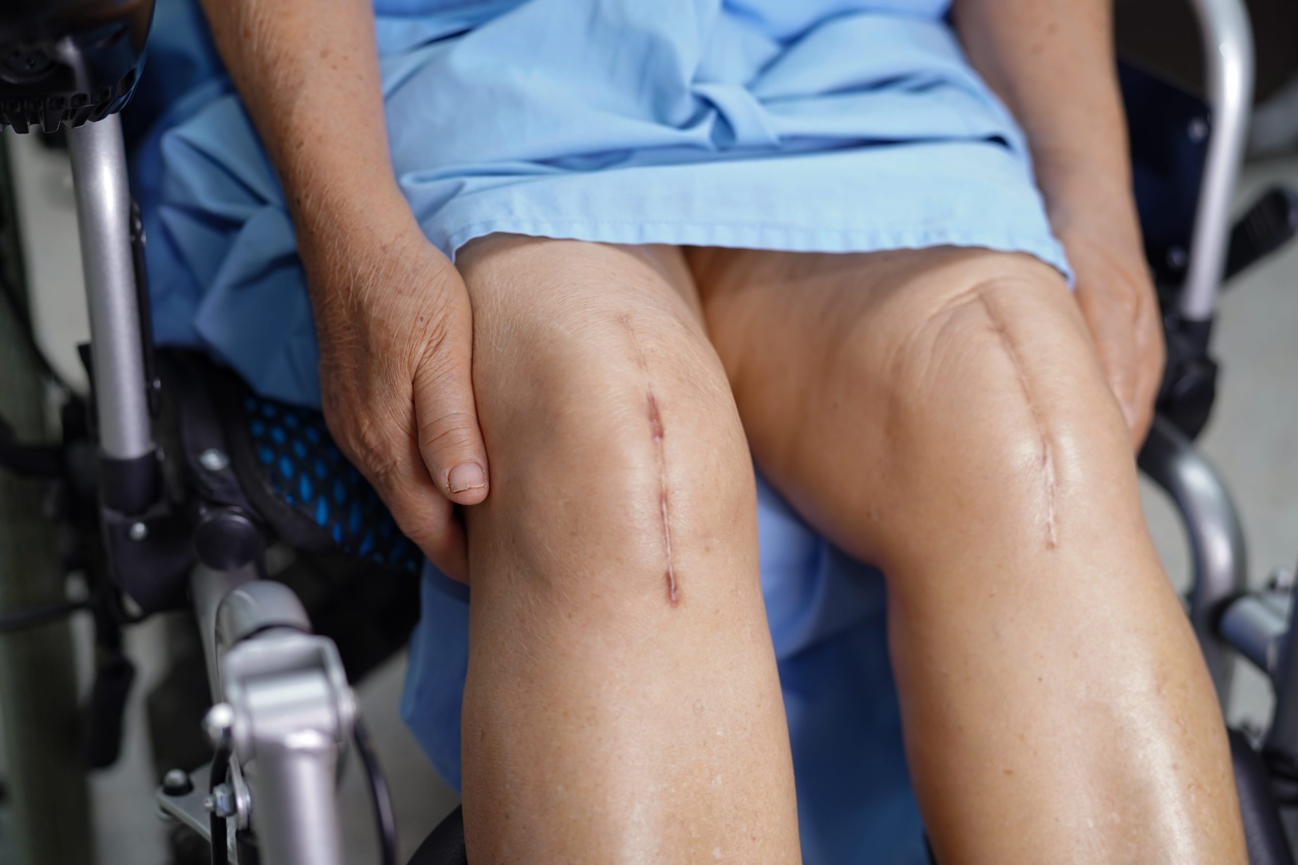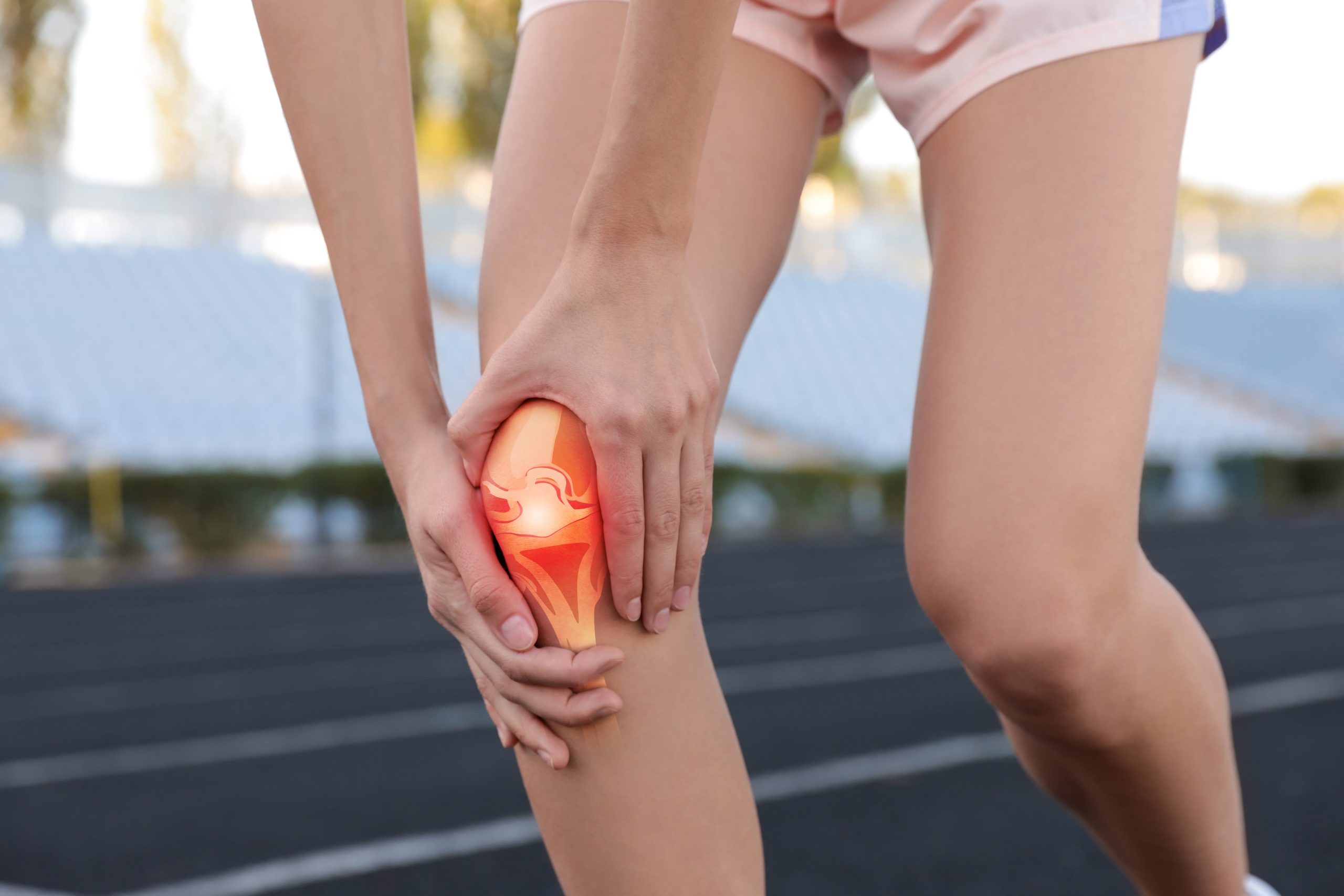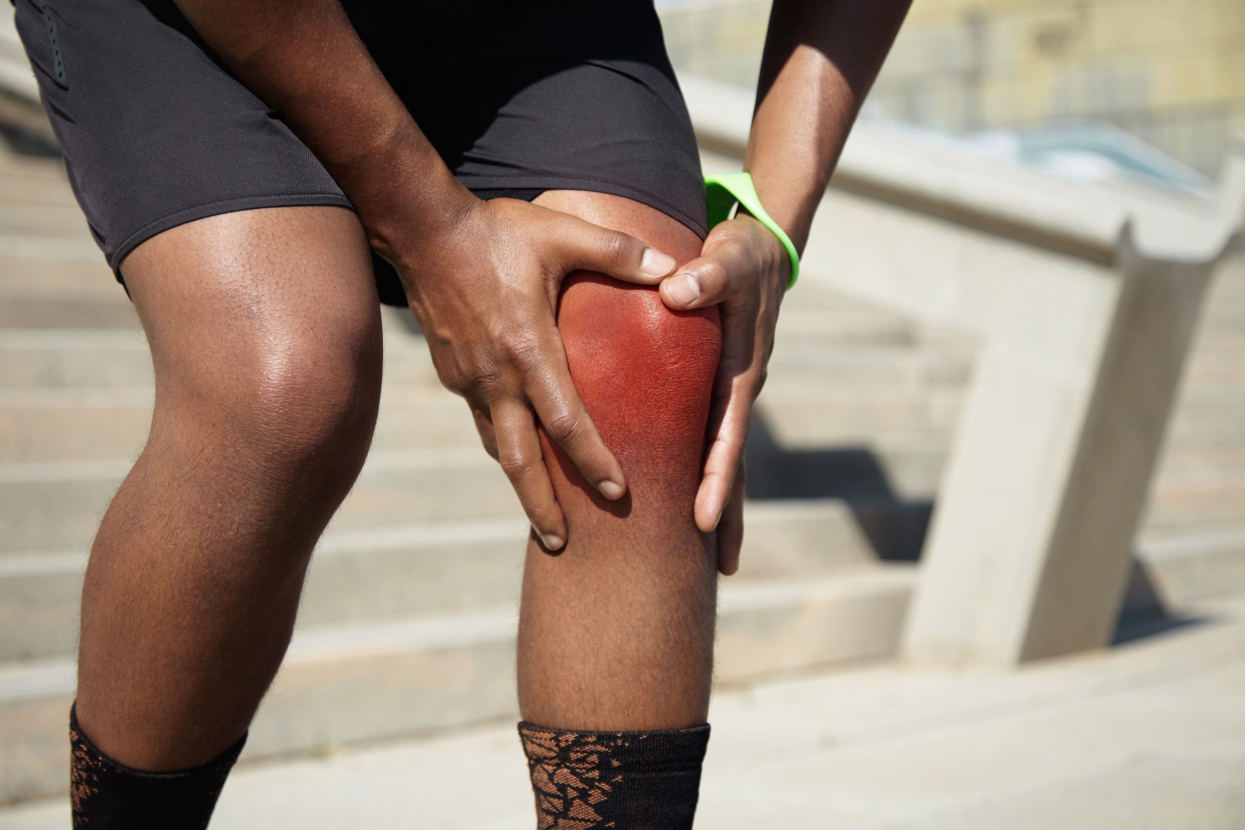
Where the swelling appears and how quickly it develops after a knee injury among the primary early clues to which structure has been damaged. The anterior cruciate ligament (ACL), medial collateral ligament (MCL), and meniscus each serve distinct mechanical functions, and understanding which structure is damaged requires recognising specific injury patterns. Each has characteristic features during the moment of injury and in the days that follow, though definitive diagnosis requires professional evaluation and often imaging studies.
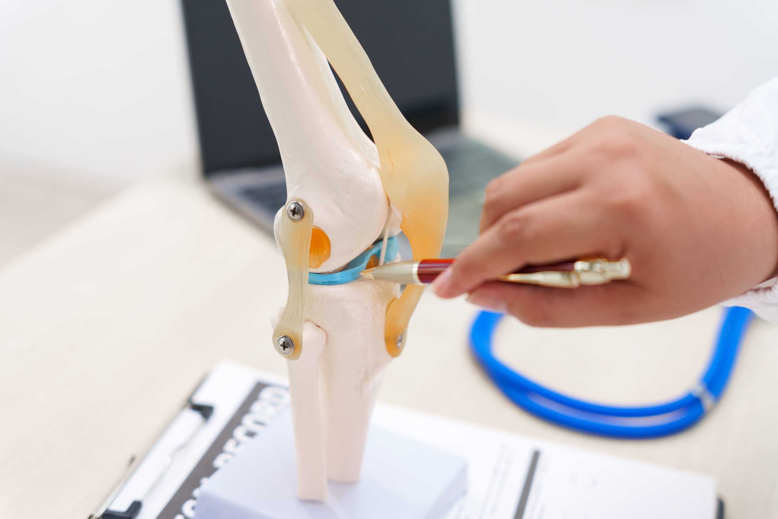
Anatomy and Function of Each Structure
The ACL runs diagonally through the centre of the knee, connecting the thighbone to the shinbone and preventing the tibia from sliding forward. This ligament also controls rotational stability, which explains why pivoting sports carry a high ACL injury risk.
The MCL sits on the inner aspect of the knee, forming a broad band that resists forces pushing the knee inward. Unlike the ACL, the MCL has access to the blood supply from surrounding tissues, which significantly influences its healing capacity.
The menisci are C-shaped cartilage pads, one medial and one lateral, that act as shock absorbers between the femur and tibia. They distribute weight across the joint surface and contribute to knee stability by deepening the relatively flat tibial plateau.
How Each Injury Typically Occurs
ACL Injury Mechanisms
ACL tears frequently occur without contact from another person. The classic mechanism involves sudden deceleration combined with a change of direction, such as planting the foot while the body rotates over a relatively straight knee. Landing from a jump with the knee extended or hyperextending the knee during an awkward step may also damage this ligament. Athletes often describe hearing or feeling a distinct pop at the moment of injury.
MCL Injury Mechanisms
MCL injuries typically result from force applied to the outer knee, pushing it inward. Direct contact with the lateral knee, such as a tackle in football or a collision during basketball, stretches or tears the lateral ligament. Skiing falls where the ski tip catches snow and forces the knee inward represent another common mechanism. The injury can also occur from planting and cutting, though less frequently than ACL injuries.
Meniscus Injury Mechanisms
Meniscal tears occur through two primary pathways. Traumatic tears happen when the knee twists while bearing weight, specifically when pivoting on a planted foot with the knee slightly bent. The trapped meniscus may tear as rotational forces exceed its structural tolerance. Degenerative tears tend to develop gradually in older adults as the cartilage loses elasticity. These may occur during minimal activity, such as squatting or rising from a chair.
Distinguishing Symptoms by Structure
ACL Tear Symptoms
Rapid, significant swelling within the first two hours may suggest ACL injury. This occurs because ACL rupture disrupts the synovial lining and surrounding vascular structures, including the synovial membrane and infrapatellar fat pad, which bleed into the joint space. Approximately 70% of patients with ACL tears experience immediate swelling from haemarthrosis. The knee may feel immediately unstable, though in some cases this sensation can be temporarily masked by pain or muscle guarding. Many patients find themselves unable to continue their activity following injury. A sensation of the knee giving way during subsequent walking or pivoting attempts indicates loss of rotational stability. Pain typically localises deep within the knee rather than along the joint line or sides.
MCL Tear Symptoms
MCL injuries may produce tenderness specifically along the inner knee, from the joint line down toward the upper shin. Swelling tends to be more localised to the medial aspect rather than generalised throughout the knee. Pain may intensify when pressure is applied to the outer knee (stressing the MCL) or when attempting to bend the knee. Unlike ACL tears, the knee may feel stable during forward-backwards motion but unstable when stressed side-to-side.
Meniscus Tear Symptoms
Meniscal tears often produce delayed swelling, developing over 24 to 48 hours rather than immediately. Joint line tenderness, meaning pain when pressing directly on the space between the femur and tibia, is characteristic. Mechanical symptoms help distinguish meniscus injuries from other conditions: catching sensations during movement, the knee locking in a bent position, or clicking with flexion and extension. Pain may typically worsen with twisting motions, squatting, or deep knee bends. Some patients can walk relatively normally after injury but experience symptoms with specific movements.
The Pop, Snap, or Tear Sensation
The audible or palpable pop most commonly occurs with ACL rupture, though it can occasionally occur with other injuries. MCL injuries more often produce a tearing sensation along the inner knee without a distinct pop. Meniscus tears may cause a click or snap, particularly during the twisting mechanism, but this is less consistently reported than the ACL pop.
Did You Know? The pop associated with ACL rupture occurs as the ligament fails under tension, releasing stored mechanical energy rapidly. The sensation travels through bone, and many patients describe it as audible even in noisy sporting environments.
Swelling Patterns and Timing
Swelling characteristics provide useful diagnostic clues. ACL tears may produce haemarthrosis, which is blood within the joint, from disruption of the synovial lining and surrounding vascular structures. This causes tense, rapid swelling that can limit knee flexion within hours. MCL injuries may create localised swelling along the medial knee that can extend down the shin, reflecting bleeding within the ligament substance and surrounding tissues. Meniscal tears tend to generate joint effusion from synovial fluid irritation, which accumulates more gradually and may fluctuate with activity levels over subsequent days.
Weight-Bearing and Walking Ability
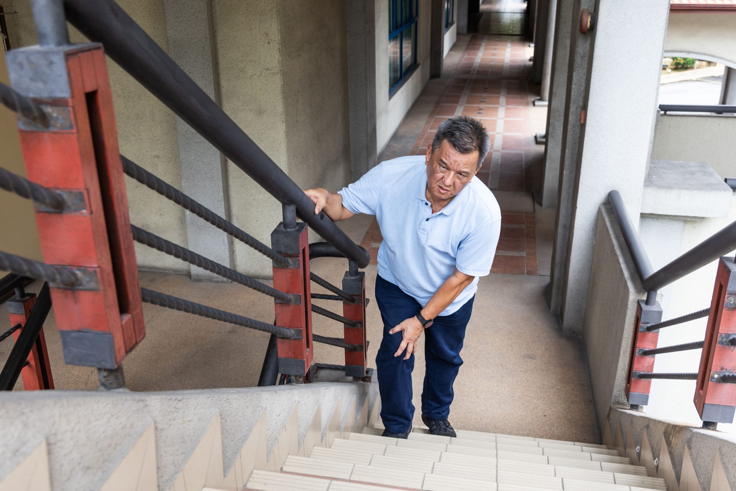
Immediately after ACL rupture, many people cannot continue their activity, though some can walk with a limp once the initial pain subsides. The knee may feel wobbly or untrustworthy during walking, particularly on uneven surfaces. MCL injuries may cause pain with each step as the ligament is stressed during normal gait, but the knee typically feels stable in the forward plane. Meniscal tears allow variable weight-bearing depending on the tear size and type. Small tears may permit near-normal walking, while large or displaced tears can cause the knee to lock, preventing full extension.
Combination Injuries
Knee trauma frequently damages multiple structures simultaneously. The classic knee injury triad involves the ACL, MCL, and a meniscus, most commonly the lateral meniscus, based on current evidence, though medial meniscus involvement is also documented. O’Donoghue originally described this triad in 1950 as involving the medial meniscus, but subsequent research has found the lateral meniscus to be more commonly injured alongside the ACL and MCL in acute sporting injuries. Combination injuries create overlapping symptom patterns that require imaging to sort out. Any injury with severe swelling, multi-directional instability, and joint line tenderness warrants evaluation for multiple structural damage.
⚠️ Important Note A knee that can be completely straightened and fully bent without mechanical blocking is less likely to have a displaced meniscal tear requiring urgent surgical intervention.
Physical Examination Tests
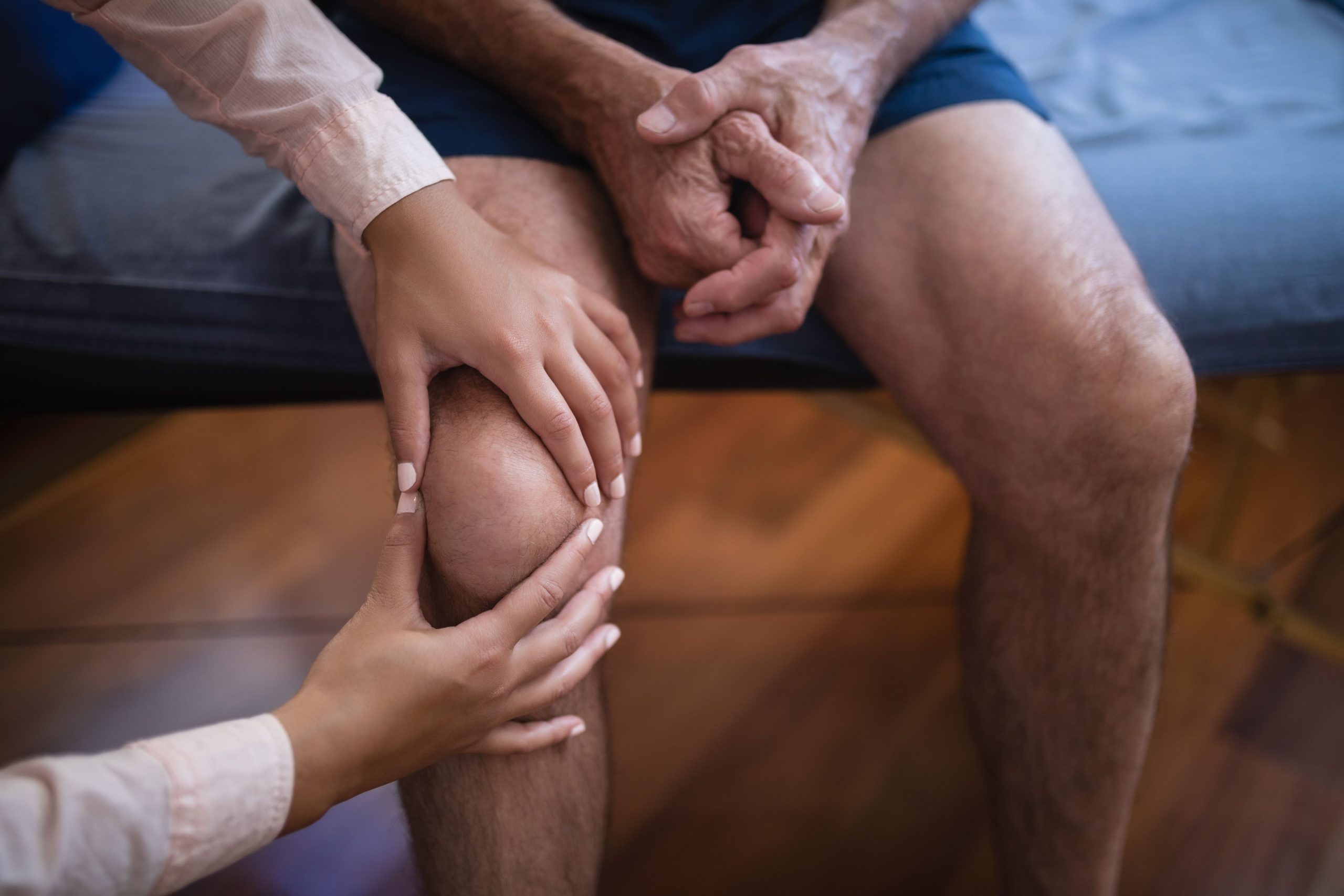
Orthopaedic specialists use specific tests to stress each structure. The Lachman test and anterior drawer assess ACL integrity by pulling the tibia forward relative to the femur. Valgus stress testing evaluates the MCL by applying an inward force to the knee at different flexion angles. McMurray’s test and joint line palpation may help identify meniscal pathology. These examinations, combined with history and symptom patterns, guide whether imaging is necessary.
When MRI Becomes Necessary
Clinical examination can identify many knee injuries, but MRI provides a more definitive structural assessment. Indications for imaging include:
- Persistent instability despite initial treatment
- Mechanical symptoms such as locking or catching
- Significant swelling that does not resolve
- Uncertainty about which structures are involved
MRI may also reveal associated injuries, including bone bruises, cartilage damage, or tears of structures not clinically suspected, that can influence treatment planning.
Recovery Timeline Differences
MCL tears generally heal without surgery because of their blood supply and healing capacity. Grade 1 sprains typically recover within one to three weeks. Grade 2 tears generally take four to six weeks. Complete Grade 3 tears may require six to twelve weeks, depending on whether other structures are also involved. ACL tears do not reliably heal spontaneously in most patients.
Non-operative management may suit lower-demand individuals, but athletes typically require reconstruction. Research indicates that returning to sport before nine months after surgery is associated with a significantly higher risk of re-injury, and most patients require nine to twelve months before returning to competitive sport. Meniscal tears vary widely. Small stable tears may settle with rest and rehabilitation over four to six weeks, while tears requiring surgical repair may need several months of protected recovery.
Practical Steps After Knee Injury
- Apply ice wrapped in a cloth for 15 to 20 minutes every few hours during the first two days to help manage swelling and discomfort.
- Use crutches if walking causes significant pain or the knee feels unstable to help reduce further stress on the joint.
- Keep the knee elevated above heart level when resting to help promote fluid drainage from the joint.
- Avoid activities that reproduce pain, catching, or giving way until a professional evaluation determines which structures are involved.
- Document how the injury occurred and how symptoms have progressed to provide your orthopaedic specialist with useful diagnostic information.
When to Seek Professional Help
- Rapid swelling within the first two hours of injury
- Sensation of the knee popping or giving way
- Inability to fully straighten or bend the knee
- Knee locking in a bent position
- Persistent instability during walking
- Pain along the joint line that does not improve over several days
- Difficulty bearing weight despite rest and ice
Commonly Asked Questions
Can I have both an ACL tear and a meniscus tear from the same injury?
Combination injuries occur frequently because the forces that rupture the ACL may simultaneously trap and tear the meniscus. MRI typically evaluates all knee structures when one major injury is suspected, specifically because associated damage is common.
Why does my knee feel stable if my ACL is torn?
Other structures, including the hamstring muscles, intact collateral ligaments, and joint capsule, may provide some stability even without an intact ACL. This compensation can mask instability during straight-line walking while failing during pivoting or cutting movements.
Do all meniscus tears require surgery?
Tear pattern, location, and patient factors determine treatment. Tears in the outer third with blood supply may heal with rest and conservative management. Degenerative tears in older adults often improve with physiotherapy. Displaced tears causing mechanical locking may typically require surgical intervention.
How accurate is self-diagnosis based on symptoms alone?
Symptom patterns provide useful guidance but cannot definitively identify which structures are damaged. Clinical examination by an orthopaedic specialist achieves reasonable accuracy, with MRI available when diagnosis remains uncertain or surgery is being considered.
Will my MCL injury need surgery?
Isolated MCL tears rarely require surgical repair regardless of severity. The ligament’s healing capacity allows many tears to recover with bracing and rehabilitation. Surgery may be considered when the MCL remains unstable after appropriate non-operative treatment or when combined with other ligament injuries requiring reconstruction.
Next Steps
Rapid swelling with instability may point toward ACL involvement; medial tenderness with side-to-side laxity may indicate MCL injury; mechanical catching with delayed swelling may suggest meniscal damage. ACL tears do not typically heal without intervention, MCL tears generally resolve with bracing and rehabilitation, and meniscal outcomes depend on tear type and location. Symptom patterns alone cannot confirm which structures are damaged. Clinical examination and imaging are required to direct appropriate treatment.
If you are experiencing knee instability, a locking sensation, joint line tenderness, or swelling that has not resolved with rest, consult our accredited orthopaedic surgeon for a clinical assessment and diagnosis.