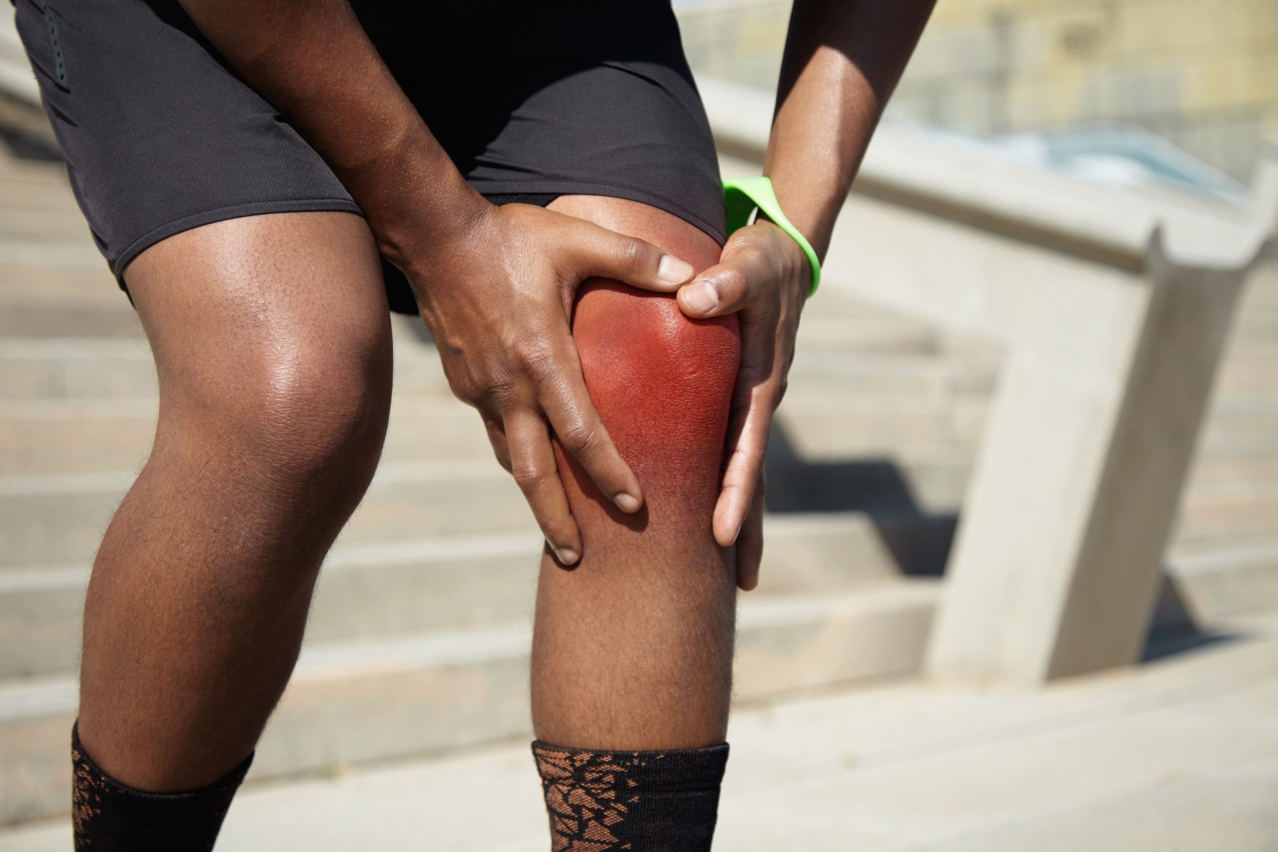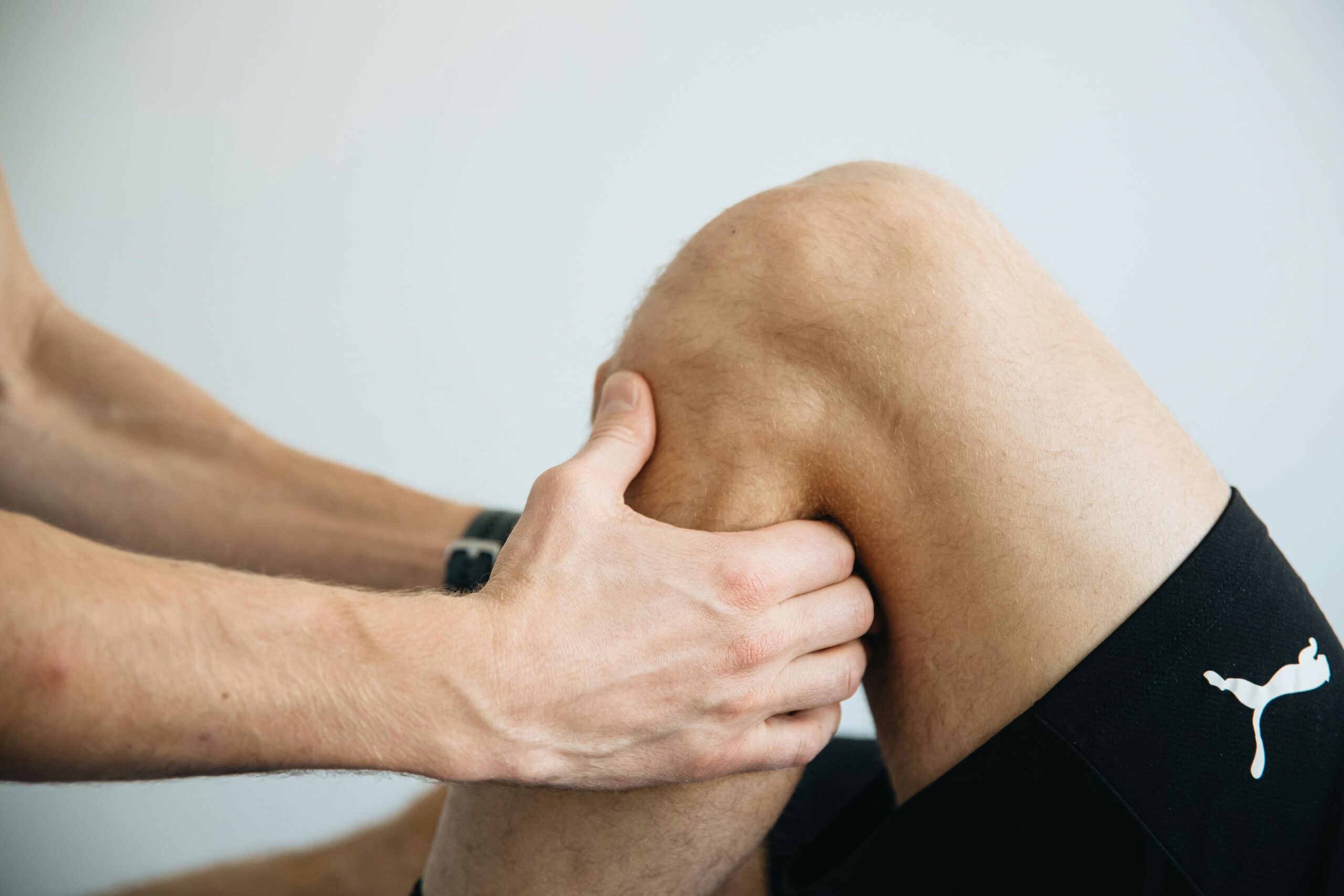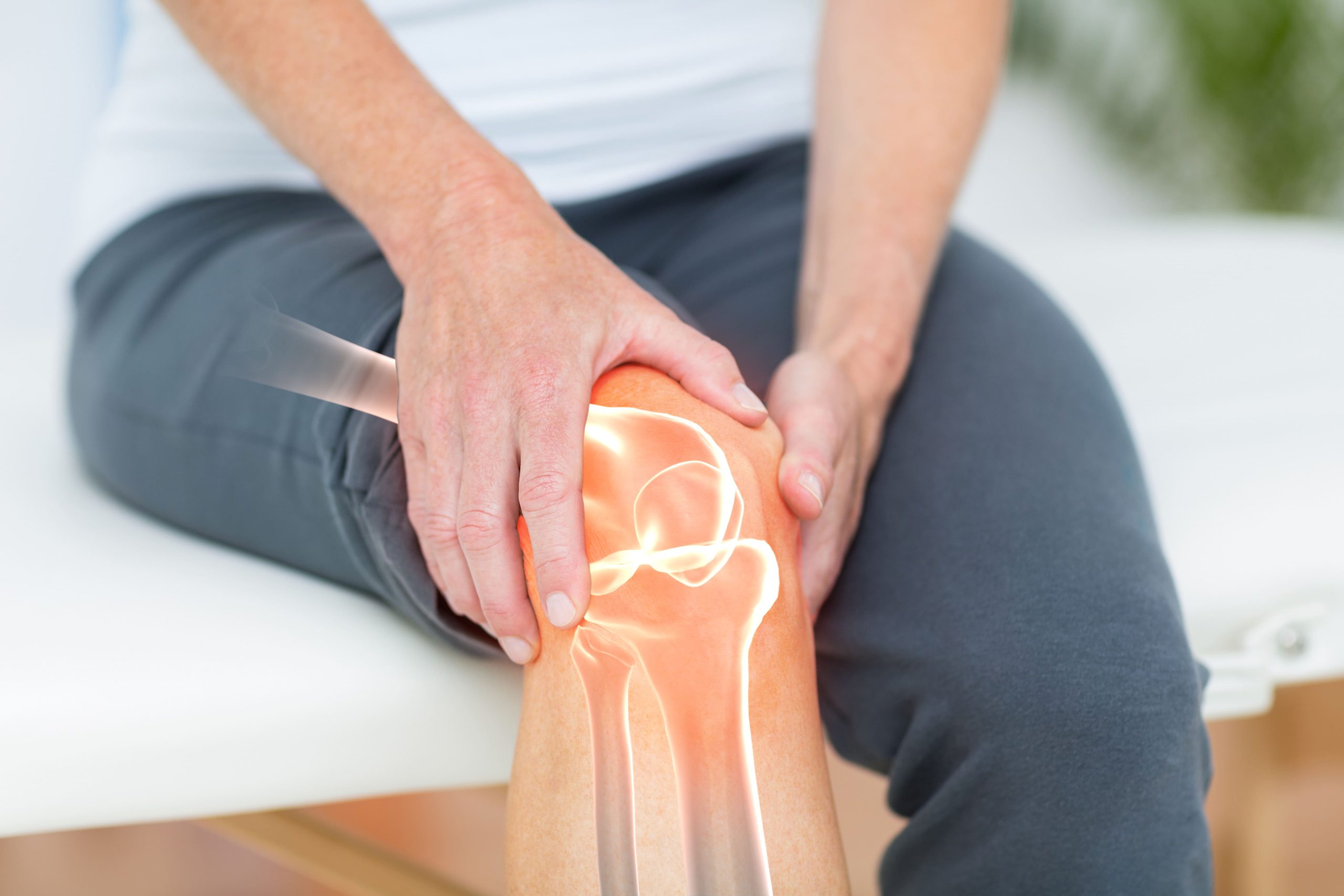
Where your knee pain is located can be an important clue about what may be causing it. The knee is a complex joint made up of ligaments, cartilage, tendons, and small fluid-filled sacs called bursae, and each of these structures can produce a different pattern of pain when irritated or injured.
Use this chart as a general guide to help you understand what your symptoms might mean, but keep in mind that an accurate diagnosis typically requires an assessment by an orthopaedic specialist, along with imaging if needed.
Quick Reference: Knee Pain Location Chart
| Location | Common Conditions | Key Symptoms |
|---|---|---|
| Front (Anterior) | Patellofemoral Pain Syndrome | Dull ache behind the kneecap; worse with stairs, squatting, prolonged sitting |
| Patellar Tendinopathy | Pain at the bottom tip of the kneecap; worse with jumping or sprinting | |
| Quadriceps Tendinopathy | Pain at the top of the kneecap; stiffness after rest; worse with deep bending | |
| Prepatellar Bursitis | Swelling directly over the kneecap; tender to touch; worse with kneeling | |
| Inner Side (Medial) |
MCL Injury | Pain along the inner knee; swelling; instability with side-to-side movement |
| Medial Meniscus Tear | Pain at the inner joint line; clicking or locking; gradual swelling | |
| Pes Anserine Bursitis | Pain 5–7 cm below the inner joint line; worse climbing stairs | |
| Medial Compartment OA | Gradual inner knee pain; morning stiffness; bow-legged appearance | |
| Outer Side (Lateral) |
IT Band Syndrome | Pain at the outer knee; begins after a predictable activity distance; resolves with rest. |
| LCL Injury | Pain along the outer knee; instability with lateral stress | |
| Lateral Meniscus Tear | Pain at the outer joint line; catching or giving way; often with other injuries | |
| Proximal Tibiofibular Joint Dysfunction | Pain below and behind the outer joint line; doesn’t fit typical patterns | |
| Back (Posterior) | Baker’s Cyst | Fullness or pressure behind the knee; may extend into the calf if ruptured |
| Hamstring Tendinopathy | Pain at the back of the knee; worse with stretching or resisted bending | |
| PCL Injury | Deep posterior pain often follows a direct blow to the front of the shin | |
| Popliteal Artery Entrapment | Calf cramping during exercise; relieved by rest; requires vascular evaluation | |
| Below the Kneecap |
Osgood-Schlatter Disease | Pain and swelling at the bony prominence below the kneecap; adolescents |
| Hoffa’s Syndrome | Sharp pain below the kneecap on either side of the tendon; worse with straightening of the knee | |
| Sinding-Larsen-Johansson | Pain at the bottom of the kneecap; adolescents during growth; resolves with maturity |
Anterior Knee Pain (Front of the Knee)
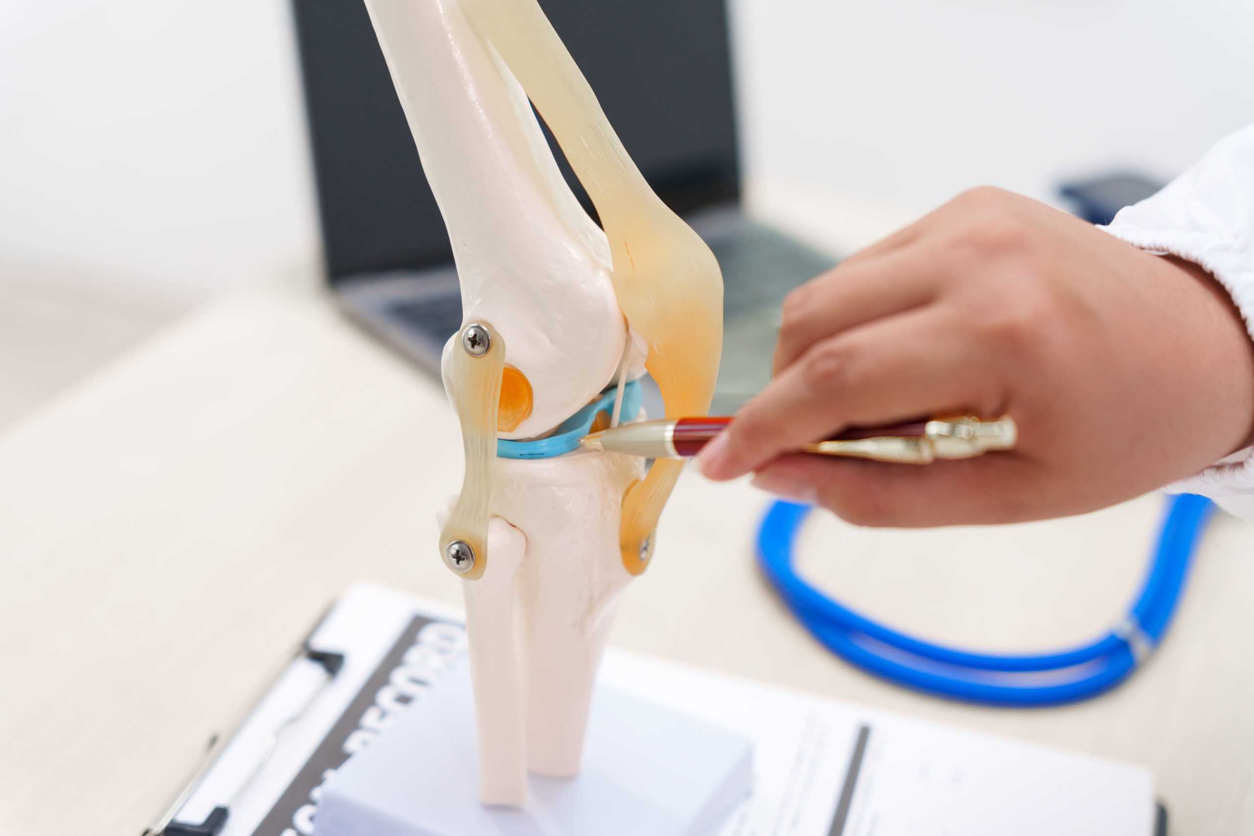
Pain at the front of the knee typically involves the kneecap (patella) and the surrounding structures that support it. The patella is a small bone embedded within the quadriceps tendon — the large tendon that connects your thigh muscles to your knee. The quadriceps tendon attaches to the top of the patella, while the patellar tendon continues downward from the bottom of the patella to your shin bone (tibia).
As you bend and straighten your knee, the patella glides smoothly along a groove at the end of the thigh bone (femur). Problems with this tracking, or with any of the tendons and sacs around the kneecap, may lead to the conditions described below.
Patellofemoral Pain Syndrome
You may feel a dull, aching pain behind or around your kneecap. The pain typically worsens with activities such as squatting, prolonged sitting, climbing stairs, and running, and it can come on gradually over time or more suddenly. Some people also notice a grinding or clicking sensation when bending the knee, or find it harder to do activities that require deep knee bending.
This is one of the most common causes of knee pain, particularly in younger, active individuals. If this sounds like your symptoms, see a physiotherapist or doctor. Strengthening the muscles around your hip and knee may help support recovery.
Patellar Tendinopathy (Jumper’s Knee)
Pain is typically felt at the bottom tip of the kneecap (the inferior pole of the patella), and is aggravated by activities that place high demand on the knee, such as those that involve jumping, landing, or sprinting. The area may feel tender when you press on it. In early or mild cases, pain may occur only during athletic activity, while more advanced cases may involve discomfort with everyday activities.
This condition is most common in athletes who play sports involving frequent jumping, such as basketball or volleyball, but it can also affect runners. Avoid pushing through pain during sport. A physiotherapist can guide you through a structured loading programme that aims to help the tendon recover.
Quadriceps Tendinopathy
This condition is characterised by activity-related pain at the front of the knee, with tenderness felt at the top edge of the kneecap (the superior border of the patella). You may also notice stiffness after periods of rest, and pain that is most pronounced with deep knee bending or during activities such as jumping and squatting. Pain when contracting the thigh muscles against resistance — such as during a leg extension exercise — is also a common feature.
This is more common in athletes and active individuals, though it can also affect people who are less active, particularly as they get older. Rest from aggravating activities and seek assessment. A physiotherapist can help you with a graded exercise plan designed to help restore tendon strength.
Prepatellar Bursitis (Housemaid’s Knee)
Prepatellar bursitis can be triggered by acute direct trauma to the knee or by repetitive pressure from frequent kneeling. It may also arise from underlying conditions such as gout, rheumatoid arthritis, or a bacterial infection.
People whose work involves a lot of kneeling — such as gardeners, plumbers, or tradespeople — are at higher risk, as are athletes in contact sports. Avoid kneeling on the affected knee. If the swelling is significant, warm, or accompanied by fever, see a doctor promptly, as an infection may need to be ruled out.
Medial Knee Pain (Inner Side)
The medial compartment contains the medial collateral ligament (MCL—a band of tissue that stabilises the inner knee), the medial meniscus, and the articulation between the femur and tibia. Pain here often relates to rotational injuries or degenerative changes.
Medial Collateral Ligament Injury
Pain localises along the inner edge of the knee, from the femoral attachment near the joint line to the tibial insertion. Injuries typically occur from valgus stress—a force pushing the knee inward while the foot remains planted. Swelling may develop along the ligament’s course. Instability with side-to-side movement indicates more severe sprains.
Medial Meniscus Tear
A medial meniscus tear causes pain concentrated at the medial joint line, the crease between the femur and tibia on the inner side. Twisting injuries commonly cause these tears. Mechanical symptoms—clicking, catching, or locking—suggest displaced meniscal tissue. Swelling typically develops gradually — often over 24 hours or more — rather than immediately, though this can vary depending on the type and severity of the tear.
Pes Anserine Bursitis
Pain and tenderness occur approximately 5–7 cm (about 2–3 inches) below the inner knee joint line, on the inner side of the shin bone. Three tendons (sartorius, gracilis, and semitendinosus) attach here. The underlying bursa can become inflamed with overuse. Pain worsens with stair climbing and may disturb sleep if lying with knees touching.
Medial Compartment Osteoarthritis
Pain develops gradually in the inner knee. It worsens with weight-bearing activities and improves with rest. Morning stiffness lasting less than half an hour is typical. You may notice the leg developing a bow-legged appearance (where the knees angle apart) as cartilage wears down on the inner side of the joint. This is called a varus deformity.
Lateral Knee Pain (Outer Side)
The lateral compartment includes the lateral collateral ligament (LCL—a band of tissue that stabilises the outer knee), lateral meniscus, and the iliotibial band (a thick band of tissue running along the outside of the thigh) where it crosses the knee. Runners and cyclists frequently experience lateral knee symptoms.
Iliotibial Band Syndrome
Pain localises to the outer knee, specifically where the iliotibial band crosses the lateral femoral condyle. The discomfort typically begins after a predictable distance or duration of activity. It resolves with rest. Tenderness concentrates at a specific point rather than diffusely along the knee’s outer edge.
Lateral Collateral Ligament Injury
Pain follows the LCL from the lateral femur to the fibular head. Injuries result from varus stress—a force pushing the knee outward. These injuries occur less frequently than MCL sprains due to the opposite leg providing protection. Instability with lateral stress testing often indicates significant damage.
Lateral Meniscus Tear
The lateral meniscus has greater natural mobility than the medial meniscus, which may offer some protection, but it remains susceptible to tears — particularly from twisting injuries and in combination with other knee ligament injuries.
Proximal Tibiofibular Joint Dysfunction
Pain localises below and slightly behind the lateral joint line where the fibula articulates with the tibia. This often-overlooked joint can become hypermobile or restricted. It may produce lateral knee symptoms that don’t fit typical patterns.
Posterior Knee Pain (Back of the Knee)
The popliteal fossa—the hollow behind the knee—contains neurovascular structures (nerves and blood vessels), the hamstring tendons, and the posterior joint capsule. Pain here requires careful evaluation to differentiate musculoskeletal from vascular causes.
Baker’s Cyst (Popliteal Cyst)
A fluid-filled swelling develops in the popliteal fossa, creating a sensation of fullness or pressure behind the knee. The cyst communicates with the joint. It often indicates underlying pathology such as meniscal tears or arthritis. If the cyst ruptures, pain and swelling may extend into the calf, mimicking deep vein thrombosis.
Hamstring Tendinopathy
Pain localises to the hamstring insertions at the back and sides of the knee. The biceps femoris attaches laterally near the fibular head. The semitendinosus and semimembranosus attach medially. Symptoms worsen with hamstring stretching and resisted knee flexion.
Posterior Cruciate Ligament Injury
The most common cause is a direct blow to the front of the shin bone while the knee is bent, such as when the knee strikes a dashboard in a motor vehicle accident or from a fall onto a bent knee. Hyperextension injuries are a less common cause.
Popliteal Artery Entrapment
Calf pain and cramping during exercise, relieved by rest, may indicate vascular compression behind the knee. This affects younger, athletic individuals. It requires vascular evaluation to differentiate from musculoskeletal causes.
Pain Below the Kneecap
Structures distal to the patella include the patellar tendon, tibial tubercle (a bony prominence where the patellar tendon attaches), and the fat pad.
Osgood-Schlatter Disease
Pain and swelling develop at the tibial tubercle, the bony prominence below the kneecap where the patellar tendon inserts. This condition commonly affects adolescents during growth spurts, particularly those involved in running and jumping sports. The tubercle may become permanently enlarged even after symptoms resolve.
Infrapatellar Fat Pad Impingement (Hoffa’s Syndrome)
Pain is felt deep to and on either side of the patellar tendon, just below the tip of the kneecap. The fat pad becomes pinched during knee extension. This produces sharp pain at the end range of straightening. Swelling around the tendon and tenderness with direct palpation are characteristic.
Sinding-Larsen-Johansson Syndrome
Unlike patellar tendinopathy — which can occur at any age — this is a growth-plate condition specific to skeletally immature adolescents and typically resolves once growth is complete.
Pain Patterns and What They Suggest
Immediate swelling Swelling that develops rapidly within the first four to six hours of injury suggests bleeding into the joint (haemarthrosis).
Gradual swelling (developing over a day or two) typically indicates meniscal injury or irritation.
Mechanical symptoms—locking, catching, or giving way—point towards structural problems like meniscal tears, loose bodies, or ligament insufficiency. Morning stiffness improving within half an hour suggests osteoarthritis (wear-and-tear arthritis). Stiffness lasting longer may indicate inflammatory conditions.
Activity-related pain that’s predictable and reproducible helps localise the source. Pain only with stair descent often indicates patellofemoral issues. Pain at a specific distance during running suggests iliotibial band syndrome. Pain with pivoting points towards meniscal or ligamentous causes.
Using a Knee Pain Location Chart Effectively
A knee pain location chart works best when you combine it with attention to symptom details:
- Mark the epicentre: Identify where pain is most intense, not just where it spreads
- Note radiation patterns: Pain may begin in one location and travel to another
- Record aggravating activities: Which movements consistently reproduce symptoms
- Track timing: When did symptoms begin, and do they follow a pattern
- Document associated findings: Swelling, instability, mechanical symptoms (such as clicking or locking), or weakness
This information creates a more complete picture for clinical evaluation. It can help focus the examination on relevant structures.
When to Seek Professional Help
- Inability to bear weight on the affected leg
- Visible deformity or significant swelling developing rapidly
- Knee locking in a bent position that won’t straighten
- Giving way or buckling during normal activities
- Pain persisting beyond two weeks despite rest and home measures
- Signs of infection: increasing redness, warmth, fever, or spreading pain
- Calf pain and swelling following knee symptoms (which requires urgent medical evaluation to rule out a blood clot (DVT).
- Numbness or tingling in the lower leg or foot
Commonly Asked Questions
Can the location of knee pain accurately predict the diagnosis?
Location provides strong diagnostic clues but may not always definitively predict the diagnosis. The knee’s compact anatomy means multiple structures occupy similar regions. A thorough examination combining location, symptom characteristics, and specific physical tests typically yields a more accurate diagnosis than location alone.
Why does my knee pain seem to move around?
Pain may genuinely affect multiple structures. Altered movement patterns from protecting one area can stress another. Additionally, referred pain from the hip or spine can manifest as knee symptoms. Persistent, shifting knee pain warrants evaluation to identify all contributing factors.
Should I use ice or heat for knee pain?
Ice can help reduce swelling and may provide temporary pain relief for acute injuries within the first couple of days. Heat generally suits chronic conditions by helping improve blood flow and reducing stiffness. If swelling is present, ice is generally preferred regardless of injury duration.
What does it mean if my knee pain is worse at night?
Night pain has several explanations: reduced distractions make pain more noticeable; inflammatory conditions often worsen at night; sleeping positions may stress affected structures. Persistent night pain, particularly if it wakes you from sleep, should be evaluated to exclude serious pathology.
Can I exercise with knee pain?
Activity modification rather than complete rest often supports appropriate outcomes. Avoid movements that reproduce sharp pain or swelling. Low-impact activities, maintaining range of motion and strength—such as swimming or cycling—frequently remain comfortable when running or jumping do not.
Next Steps
Pain location, symptom timing, aggravating activities, and associated findings—such as swelling, mechanical symptoms, or instability—provide the most clinically useful picture. Symptoms persisting beyond 2 weeks, mechanical issues such as locking or giving way, or any functional limitation warrant a professional assessment to establish an accurate diagnosis and an appropriate treatment plan.
If you are experiencing persistent knee pain, joint locking, giving way, or swelling that limits your daily activities or sports, an orthopaedic specialist can evaluate your condition through examination and appropriate imaging to determine the source and discuss treatment options.