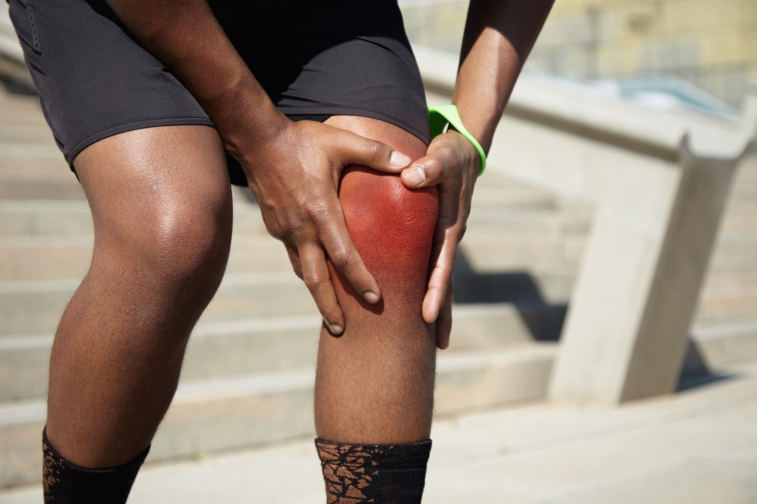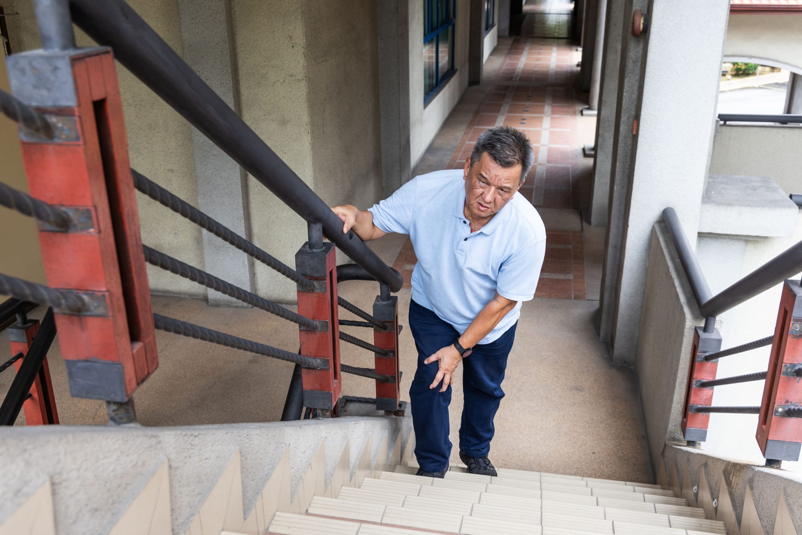
The fibrocartilage cushioning your knee joint can tear gradually over years without any single incident causing it, which is what sets degenerative meniscus tears apart from the acute tears seen in sport. Rather than a pivoting injury, an awkward fall, or any identifiable moment of damage, the tissue loses water content and structural integrity over decades until ordinary movements like climbing stairs, squatting, or stepping off a curb become consistently painful.
Someone with this gradual form of cartilage breakdown faces different decisions from a young athlete with an acute sports tear, with surgery becoming one option among several rather than an automatic recommendation.
How Degenerative Meniscus Tears Develop
Age-related changes in meniscal tissue gradually reduce its resilience until everyday movements become enough to cause damage.
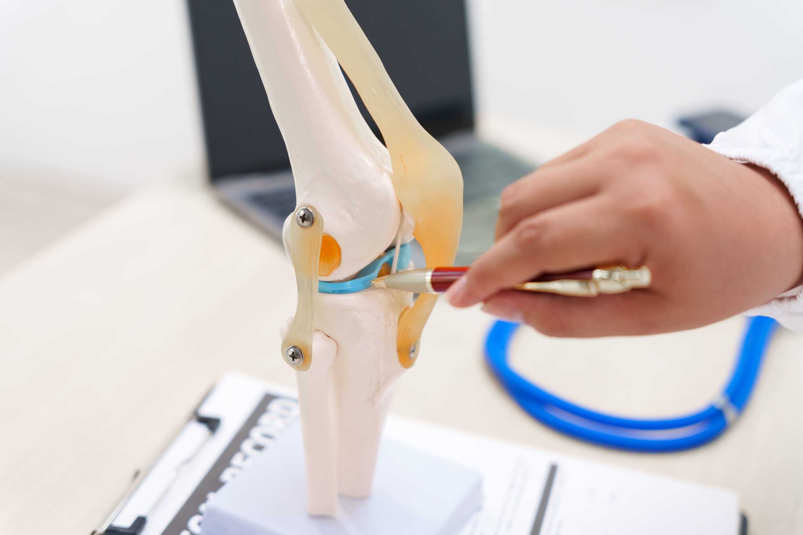
- Structural deterioration with age: Meniscal cartilage contains collagen fibres arranged in specific patterns that give it strength and flexibility. Over decades, these fibres become disorganised, and the tissue loses proteoglycans, the molecules that attract water and keep the meniscus plump and resilient.
- Everyday triggers: Because the tissue is weakened, activities that wouldn’t damage a healthy meniscus, such as a deep squat, an unexpected step off a curb, or twisting while carrying groceries, can cause the brittle cartilage to fray or split. Many people experience their first symptoms during such ordinary movements, leading to confusion about when the problem actually started.
- Which meniscus is more affected: The medial meniscus (inner side of the knee) sustains more degenerative damage than the lateral meniscus (outer side), reflecting the greater load the inner compartment bears during walking and standing.
- Most common tear pattern: Horizontal cleavage tears, where the meniscus splits along its horizontal plane, represent the most common degenerative pattern and are distinct from the vertical tears typically seen in younger patients with acute injuries.
Recognising Symptoms of Gradual Meniscus Damage
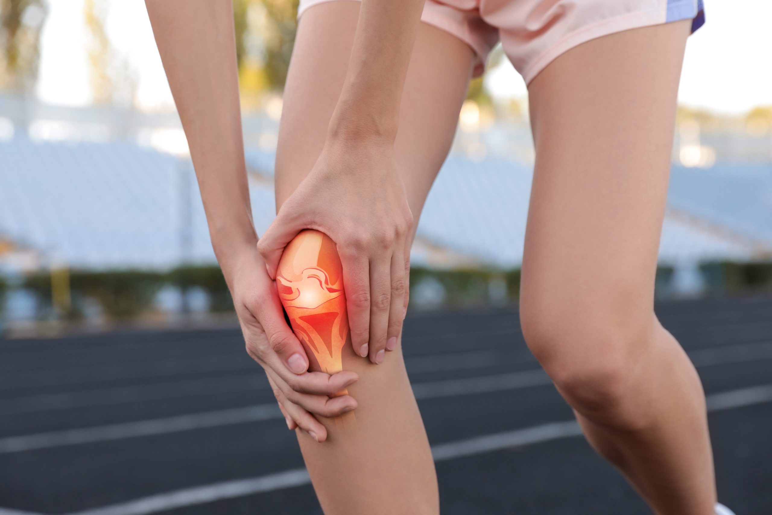
Pain along the joint line, the space between your thighbone and shinbone, represents the hallmark symptom. This discomfort typically localises to one side of the knee, corresponding to which meniscus is affected. Pressing directly on this area often reproduces the pain.
Mechanical symptoms distinguish meniscus problems from other causes of knee pain:
- Catching: A sensation that something snags inside the joint during movement
- Locking: Temporary inability to fully straighten the knee, as if something blocks extension
- Giving way: Sudden instability, often when changing direction or on uneven surfaces
- Clicking or popping: Audible or palpable sounds with knee movement
Swelling tends to be mild and intermittent rather than the dramatic swelling seen with acute injuries. The knee may feel puffy after activity and settle overnight. Morning stiffness lasting under 30 minutes is common; longer duration suggests inflammatory arthritis rather than isolated meniscal damage.
Pain patterns vary considerably. Some people experience constant low-grade aching. Others have pain-free intervals punctuated by flare-ups triggered by specific activities. Squatting, kneeling, and twisting movements typically aggravate symptoms more than straight-line walking.
The Connection Between Meniscus Tears and Osteoarthritis
Degenerative meniscus tears rarely exist in isolation. MRI studies of adults over 50 frequently reveal meniscal abnormalities in people without any knee symptoms; the changes are so common they’re considered part of normal ageing. When symptoms do develop, they often reflect a combination of meniscal damage, cartilage wear, and early osteoarthritis.
This overlap complicates treatment decisions. A tear visible on MRI may or may not be the actual source of someone’s pain. The arthritic changes in the surrounding joint surfaces might contribute equally or more to symptoms. Addressing only the meniscus while ignoring broader knee arthritis and joint health rarely provides lasting relief.
The meniscus and articular cartilage exist in a relationship where damage to one accelerates the deterioration of the other. A torn meniscus no longer distributes load evenly, increasing stress on specific areas of cartilage. Damaged cartilage alters joint mechanics, increasing meniscal stress. This cycle explains why treatment must consider the entire joint rather than focusing solely on the tear.
Did You Know?
The meniscus has limited blood supply, with only the outer third receiving direct blood flow. This “red zone” can potentially heal, while tears in the inner “white zone” lack the biological resources for spontaneous repair, a factor in surgical planning.
Diagnostic Evaluation Process
Clinical examination, targeted imaging, and occasionally diagnostic injection combine to build a complete picture of meniscal damage and guide treatment decisions.
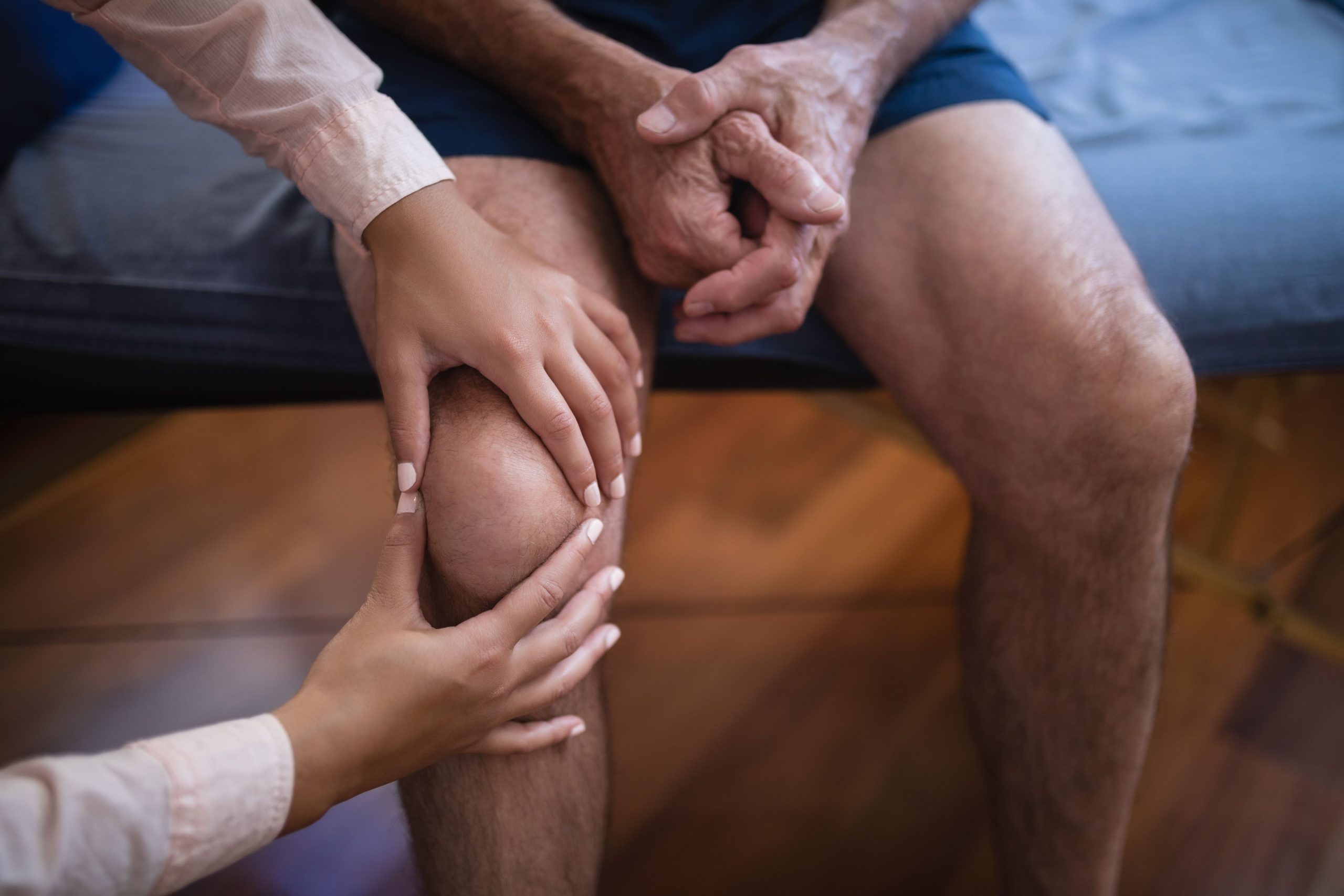
- Clinical examination: Your orthopaedic surgeon assesses swelling patterns, range of motion, and joint line tenderness and performs specific manoeuvres that stress the meniscus. The McMurray test, in which the examiner first places the knee in full flexion, then applies tibial rotation (external rotation to assess the medial meniscus, internal rotation for the lateral meniscus) before passively extending the knee, may reproduce pain or a palpable click when meniscal tissue is caught between joint surfaces.
- X-rays: These cannot visualise the meniscus directly but reveal important information about joint space narrowing, bone spurs, and alignment, all relevant to treatment planning. Standing X-rays, taken while you bear weight, show the functional state of the joint more accurately than images taken lying down.
- MRI: This provides detailed images of soft tissue structures, including both menisci, ligaments, and articular cartilage, showing tear location, pattern, and size whilst also revealing associated findings like cartilage damage, bone marrow lesions, and cysts. MRI findings must be interpreted alongside clinical symptoms, as not every visible tear requires treatment.
- Diagnostic injection: Occasionally, a local anaesthetic injection into the joint helps determine whether the meniscus is the primary pain source. Temporary relief following injection suggests intra-articular pathology, though it doesn’t distinguish between meniscal and other causes.
Non-Surgical Treatment Approaches
Initial management typically involves conservative measures regardless of MRI findings. Many degenerative tears improve substantially without surgery, particularly when symptoms are recent or mechanical symptoms are absent.
Activity modification reduces stress on the damaged meniscus. This doesn’t mean complete rest but rather avoiding activities that consistently provoke pain. Temporarily substituting cycling or swimming for running, using a lift instead of stairs, or breaking prolonged standing into shorter intervals allows symptoms to settle.
Physical therapy addresses muscle weakness and movement patterns that increase joint stress. Quadriceps and hip strengthening reduce the load transmitted through the meniscus. Balance training improves joint stability. Manual therapy techniques may improve range of motion and reduce stiffness.
Anti-inflammatory medications provide symptomatic relief during flare-ups. Short courses of oral NSAIDs or topical preparations reduce pain and swelling. These medications treat symptoms rather than the underlying problem and work best as part of a broader management plan.
Corticosteroid injections offer temporary relief when oral medications prove insufficient. A single injection into the joint can reduce inflammation and pain for weeks to months. Repeated injections are limited due to potential effects on cartilage health with long-term use.
Hyaluronic acid injections aim to improve joint lubrication. Evidence for their effectiveness remains mixed, but some patients report meaningful symptom reduction. These injections are typically considered when other conservative measures provide inadequate relief.
When Degenerative Meniscus Tear Surgery Makes Sense
The role of surgery for degenerative tears has evolved substantially based on research comparing surgical and non-surgical outcomes. Current evidence suggests that arthroscopic partial meniscectomy, removing the torn portion of the meniscus, provides limited benefit over comprehensive physical therapy for most patients with degenerative tears.
However, specific situations exist where surgery offers clear advantages:
True mechanical locking that prevents full knee extension and doesn’t resolve with conservative care typically requires surgical removal of the obstructing tissue. A displaced bucket-handle tear fragment blocking joint movement won’t improve without intervention.
Failed conservative treatment after adequate duration, typically several months of appropriate therapy, may indicate surgery. “Adequate” means actually completing prescribed exercises, not simply waiting whilst avoiding rehabilitation.
Specific tear patterns may benefit from surgery even in degenerative knees. Large flap tears that catch consistently, or tears with unstable fragments causing recurrent locking, respond better to surgical treatment than small, stable degenerative tears.
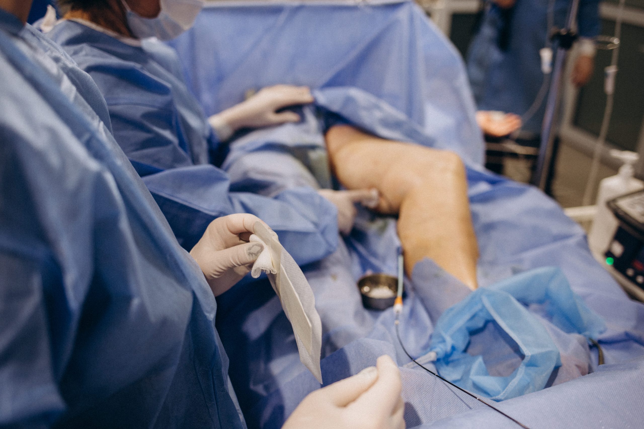
The procedure involves knee arthroscopy, inserting a small camera and instruments through two or three small incisions. The surgeon removes only the damaged, unstable tissue whilst preserving as much healthy meniscus as possible. Complete meniscus removal is avoided due to the protective role the meniscus plays in preventing arthritis progression.
Important Note:
Meniscus repair, stitching the torn edges back together, is rarely possible for degenerative tears. The tissue quality is usually insufficient to hold sutures, and most degenerative tears occur in areas without the blood supply needed for healing.
Recovery and Rehabilitation After Surgery
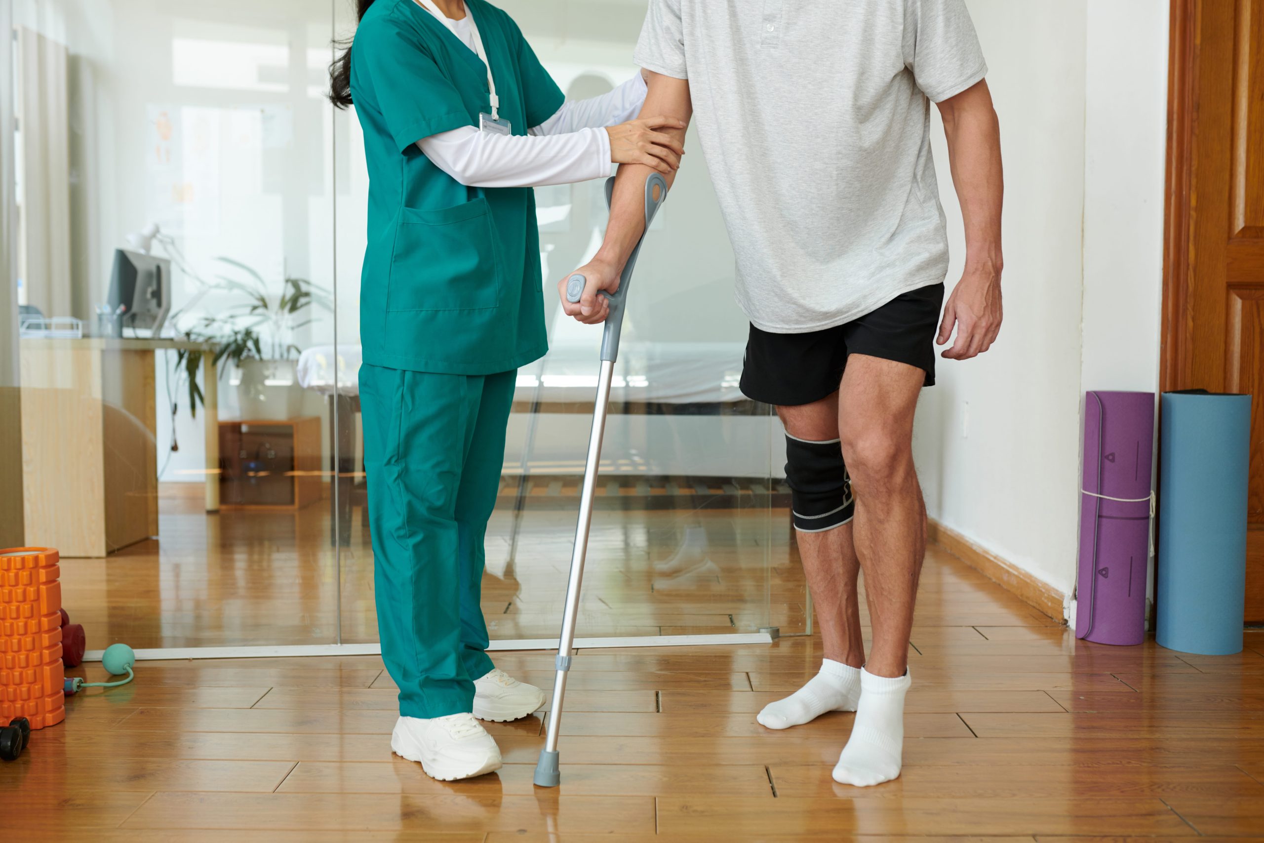
Arthroscopic meniscectomy typically allows weight-bearing immediately, though some surgeons prefer a brief period of protected walking with crutches. Initial recovery focuses on controlling swelling, restoring range of motion, and gradually rebuilding muscle strength.
Most patients return to desk work within days and physically demanding occupations within weeks to months, depending on job requirements. Full recovery, meaning return to all activities without restriction, generally occurs over one to three months.
Rehabilitation plays an important role regardless of surgical or non-surgical treatment.
Post-operative physical therapy follows similar principles to conservative treatment: quadriceps strengthening, restoration of balance and proprioception, and gradual return to functional activities. Patients who skip or abbreviate rehabilitation typically have worse outcomes than those who complete their prescribed programmes.
Long-Term Joint Health Considerations
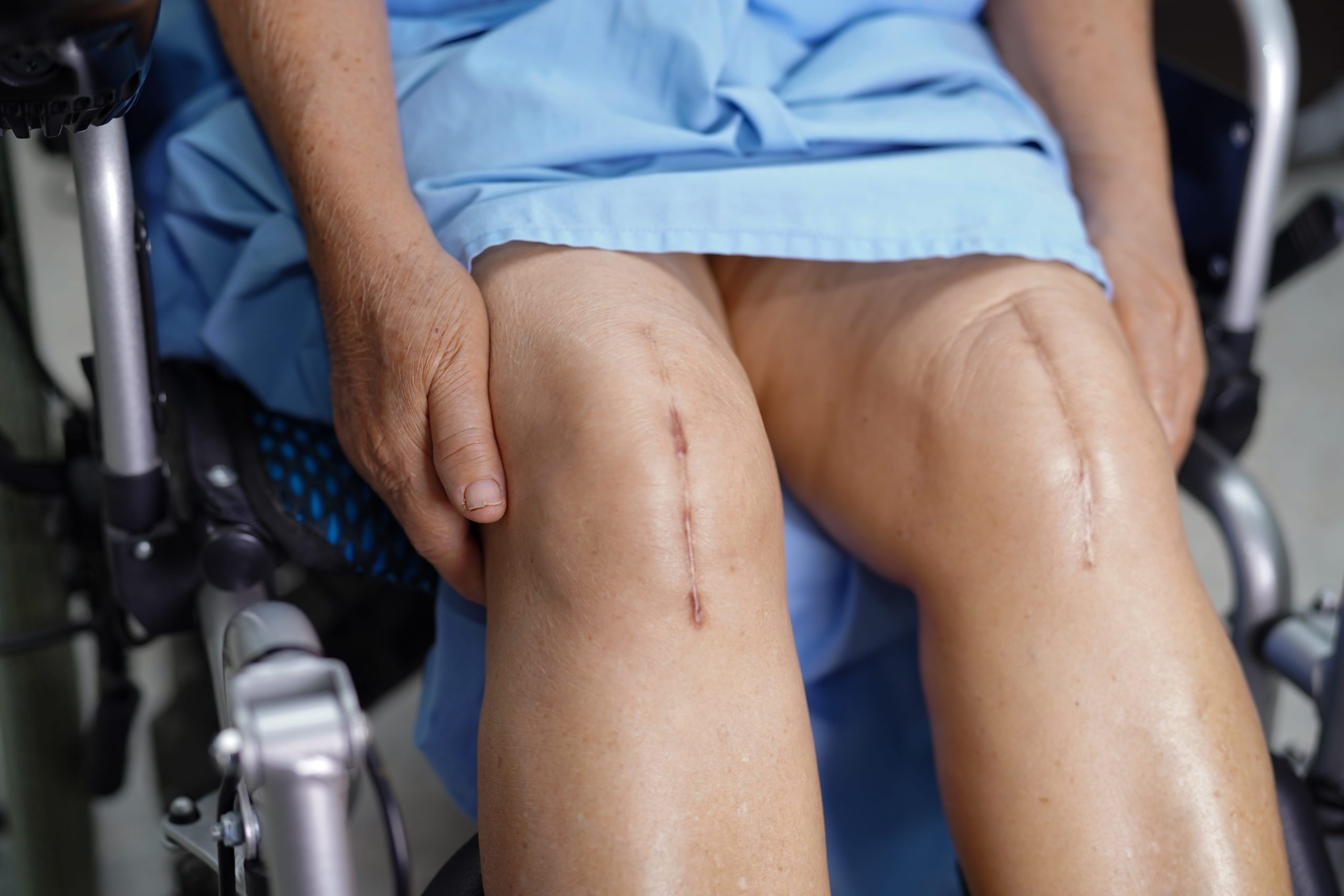
Meniscus removal, even partial, changes knee biomechanics permanently. The remaining joint surfaces bear increased stress in areas previously protected by the excised tissue. Over decades, this accelerates cartilage wear in the affected compartment.
This long-term consideration influences the conservative approach to degenerative tears. If symptoms can be adequately managed without surgery, the meniscal tissue, even though damaged, continues providing some protective function. Removing it provides a short-term benefit but potentially accelerates the arthritic process.
Ongoing joint health maintenance involves maintaining a healthy weight, staying physically active with joint-friendly activities, and addressing any lower limb alignment or muscle imbalance issues. Regular physical activity actually protects joints better than excessive rest, as movement nourishes cartilage and maintains supporting muscle strength.
Managing Daily Activities With Meniscus Damage
Modify impact activities by choosing lower-impact alternatives during symptomatic periods. Swimming, cycling, and elliptical training maintain cardiovascular fitness whilst reducing joint stress.
Strengthen supporting muscles through regular exercise targeting the quadriceps, hamstrings, and hip abductors. Stronger muscles absorb more shock before it reaches the joint.
Use appropriate footwear with adequate cushioning and support. Worn-out shoes increase joint loading with every step.
Manage body weight within healthy ranges, as additional weight multiplies the load through the knee during walking and especially stair climbing.
Apply ice after aggravating activities to reduce inflammation. Ice packs for 15 to 20 minutes help settle flare-ups.
When to Seek Professional Help
- Knee pain persisting beyond several weeks despite rest and over-the-counter medications
- Locking or catching that prevents normal knee movement
- Significant swelling that develops after activity
- Giving way or instability during walking
- Pain that interferes with sleep or daily activities
- Knee pain accompanied by visible deformity or inability to bear weight
Commonly Asked Questions
Can a degenerative meniscus tear heal on its own?
The inner portion of the meniscus lacks the blood supply needed for tissue healing, so the tear itself doesn’t repair spontaneously. However, symptoms often improve substantially as inflammation settles and surrounding structures adapt. Many people with MRI-confirmed tears become asymptomatic without surgical intervention.
How do I know if I need surgery or can manage without it?
Mechanical symptoms, particularly true locking where you cannot straighten your knee, suggest surgery may be necessary. Pain and swelling alone, even if significant, often respond to conservative treatment. A trial of structured physical therapy typically clarifies whether surgery is needed.
Will my knee develop arthritis after a degenerative meniscus tear?
Degenerative tears frequently coexist with early arthritic changes. The tear and arthritis share common causes related to accumulated joint stress over time. Whether treated surgically or conservatively, maintaining muscle strength, a healthy weight, and regular activity helps slow arthritis progression.
How long should I try physical therapy before considering surgery?
A meaningful trial typically requires two to three months of consistent, appropriate rehabilitation. Simply attending sessions without performing home exercises or waiting passively without active treatment doesn’t constitute an adequate conservative trial.
Is arthroscopic surgery a minor procedure?
Whilst arthroscopy is less invasive than open surgery, it remains a genuine surgical procedure with associated risks including infection, blood clots, and anaesthesia complications. Recovery requires active rehabilitation. Understanding this helps set appropriate expectations.
Next Steps
A degenerative meniscus tear visible on an MRI does not automatically require surgery; individual symptoms, mechanical findings, and response to rehabilitation determine the appropriate course. A structured trial of physical therapy lasting two to three months is the standard first step for most patients. Surgery is typically reserved for true mechanical locking, large unstable tear patterns, or the failure of adequate conservative treatment. Partial meniscectomy, when indicated, aims to preserve as much stable meniscal tissue as possible to reduce long-term arthritic risk.
If you are experiencing persistent knee pain accompanied by catching, mechanical locking, or joint instability, a comprehensive clinical examination and targeted imaging by an accredited orthopaedic specialist can help clarify whether a conservative management plan or an orthopaedic consultation is the most appropriate next step for your condition.