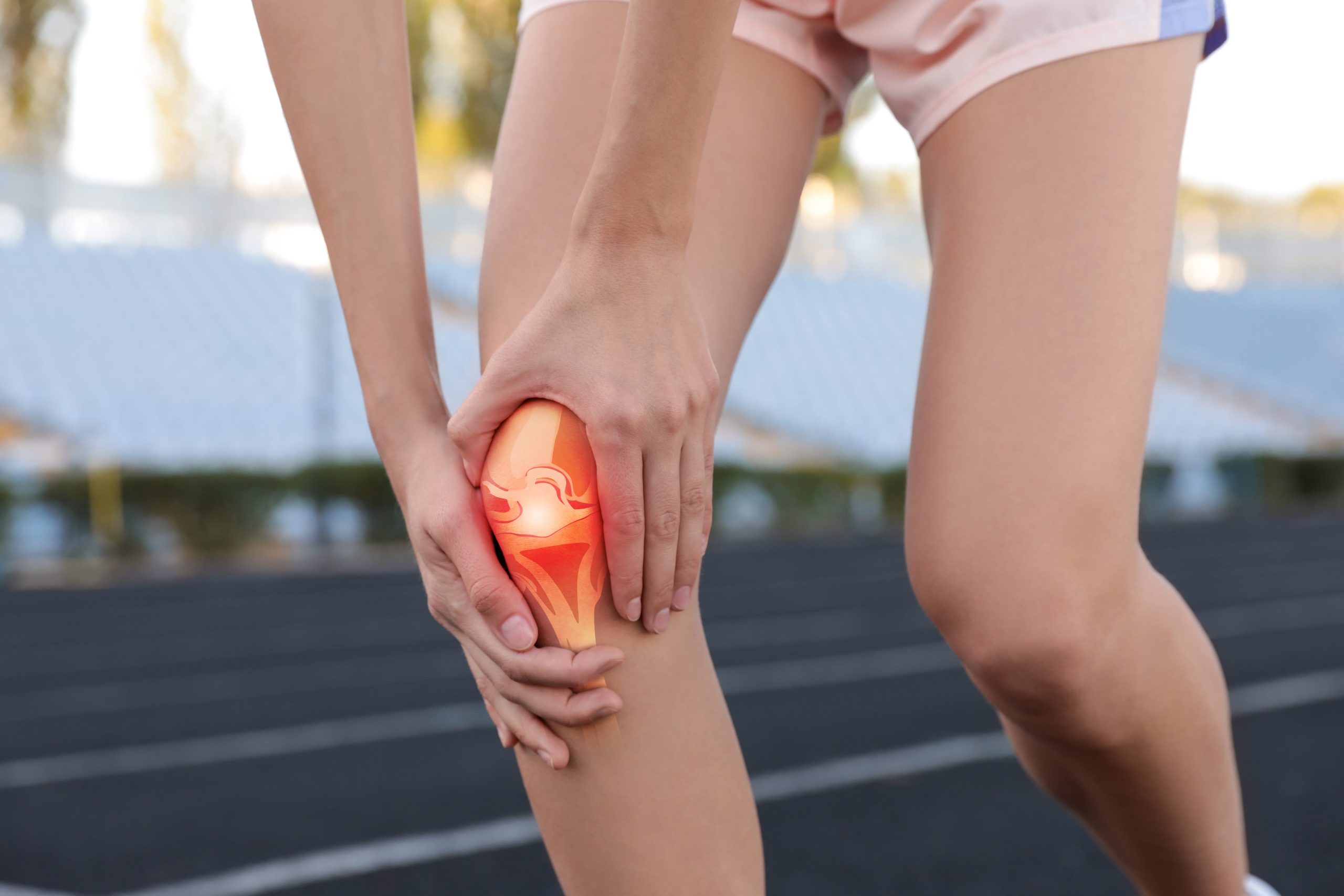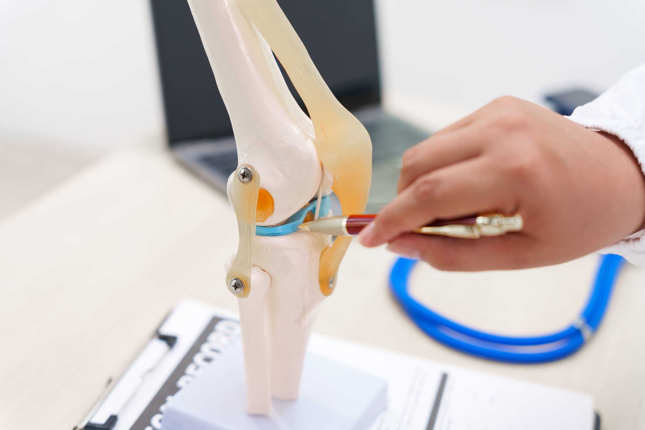
The medial and lateral menisci are two C-shaped cartilage structures in the knee that serve as shock absorbers, distributing weight across the joint surface, but they differ in structure, movement patterns, and injury characteristics. The medial meniscus sits on the inner side of the knee and remains relatively fixed to the joint capsule, while the lateral meniscus on the outer side moves more freely during knee motion. These anatomical differences directly influence how each meniscus tears, heals, and responds to treatment.
Anatomical Differences That Matter
The medial and lateral menisci differ in size, blood supply, and attachment, and these differences directly influence injury risk, healing potential, and treatment planning.
- Tibial plateau coverage: The medial meniscus covers approximately 50 to 60% of the medial tibial plateau, while the lateral meniscus covers roughly 70 to 80% of the lateral plateau, meaning lateral meniscus injuries may affect a proportionally larger weight-bearing area.
- Blood supply zones: Both menisci are divided into three vascular zones. The outer third (red zone) receives adequate blood flow and may heal more reliably. The middle third (red-white zone) has limited vascularity. The inner third (white zone) lacks direct blood supply, making tears in this region less likely to heal without intervention.
- Medial meniscus attachment: The medial meniscus attaches firmly to the joint capsule along its entire periphery and connects to the deep fibres of the medial collateral ligament, limiting its ability to move away from compressive or rotational forces.
- Lateral meniscus attachment: The lateral meniscus has looser capsular attachments and connects to the popliteus tendon, allowing it to shift posteriorly during knee flexion, a protective mechanism that may partially account for its lower overall injury rate.
How Each Meniscus Tear Differently
Medial meniscus tears commonly occur during weight-bearing activities combined with twisting motions. Pivoting on a planted foot while changing direction creates shearing forces that the fixed medial meniscus cannot easily escape. Sports involving cutting movements, sudden stops, and direction changes may frequently produce these injuries.
The lateral meniscus typically tears during deep squatting activities or hyperflexion injuries. Its posterior horn becomes compressed between the femoral condyle and tibial plateau at extreme flexion angles. Athletes in sports requiring deep knee bends, such as weightlifting, certain martial arts positions, or catcher stances in baseball, may face a higher risk of lateral meniscus injury.
Degenerative tears affect both menisci but follow different patterns. The medial meniscus tends to develop horizontal cleavage tears and complex degenerative patterns more frequently with age. The lateral meniscus shows a higher proportion of horizontal tears extending to the popliteal hiatus region.
Did You Know? The lateral meniscus transmits approximately 70% of the load in the outer knee compartment, compared to roughly 50% for the medial meniscus on the inner side, making lateral meniscus preservation an important consideration for long-term joint health.
Recognising Symptoms of Each Type
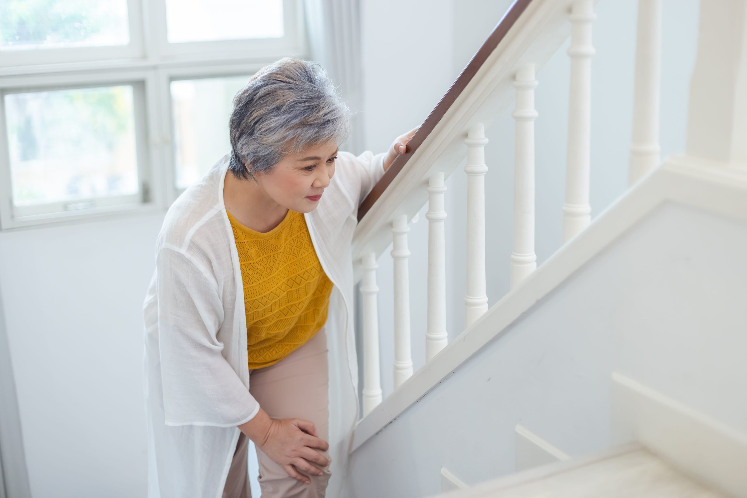
Medial meniscus tears may produce pain along the inner knee joint line, often worsening with deep knee bends or when rising from a seated position. Patients frequently report a catching sensation when walking or climbing stairs. Swelling typically develops gradually over several hours rather than immediately after injury.
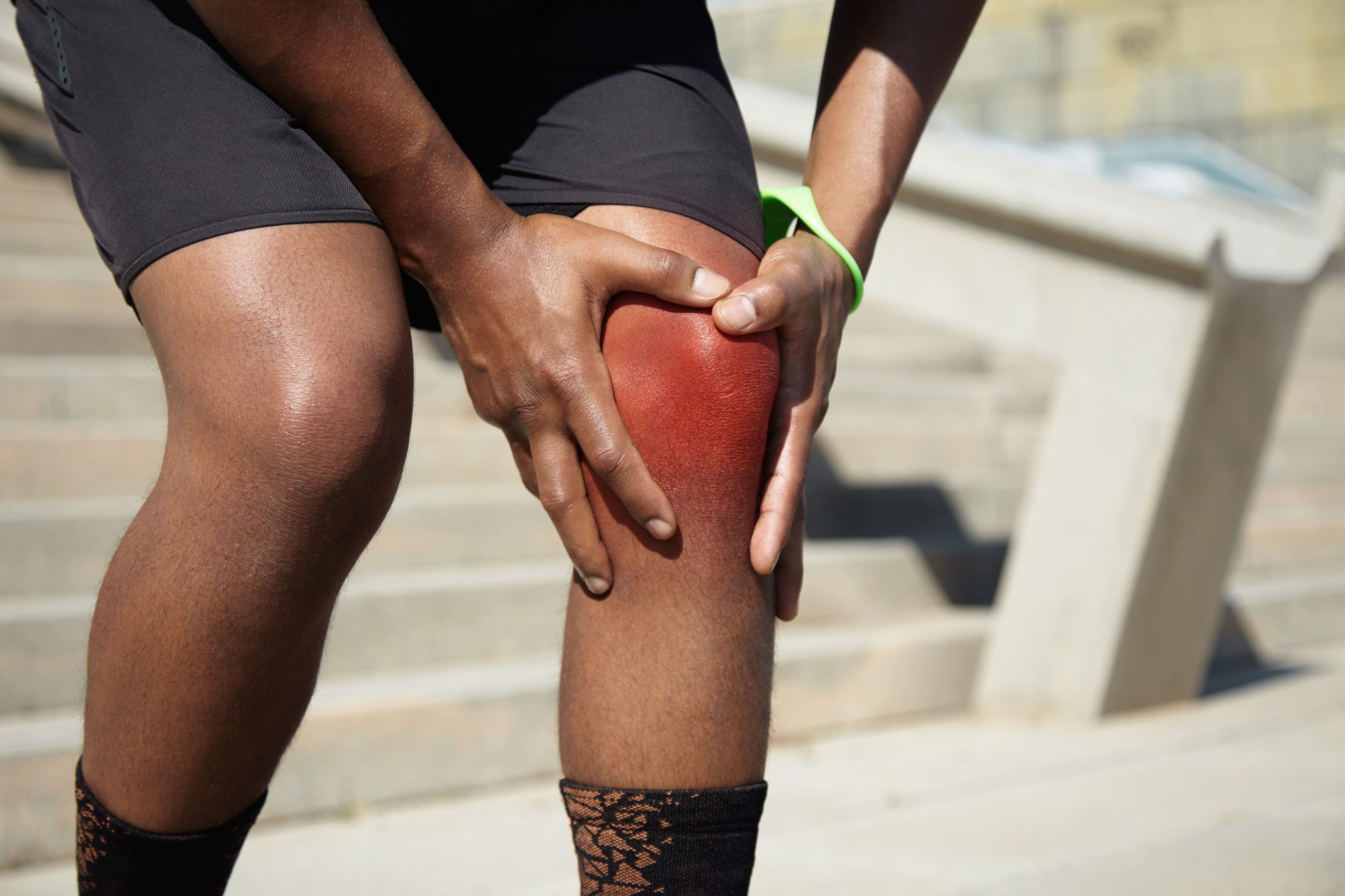
Lateral meniscus tears may cause pain along the outer joint line that can radiate toward the back of the knee. The catching or locking sensation often occurs during the final degrees of knee straightening. Some patients notice symptoms worsen when walking on uneven surfaces or during lateral movements.
Both tear types can cause:
- Joint line tenderness at the specific location
- Swelling that fluctuates with activity levels
- Difficulty achieving full knee extension
- A sensation of the knee giving way
- Pain with squatting or twisting movements
The McMurray test and Thessaly test may help clinicians differentiate between medial and lateral involvement during physical examination, though MRI provides detailed imaging to help confirm the diagnosis and reveals tear configuration.
Tear Patterns and Their Significance
Vertical longitudinal tears run parallel to the meniscus fibres and occur more commonly in younger patients following acute trauma. When these tears extend significantly, they can create a bucket handle configuration where the torn segment may flip into the joint centre, potentially causing mechanical locking.
Radial tears cut perpendicular to the fibre direction and may disrupt the hoop stress mechanism that allows the meniscus to distribute load. These tears can compromise meniscal function significantly, often presenting different clinical challenges compared to longitudinal tears of similar size.
Horizontal cleavage tears split the meniscus into upper and lower layers, typically developing gradually in patients over 40. These tears may produce meniscal cysts when synovial fluid tracks through the cleavage plane to the joint periphery.
Complex tears combine multiple patterns and usually indicate degenerative changes. Treatment options may become more limited as tear complexity increases.
⚠️ Important Note: A locked knee, meaning an inability to fully straighten the joint, warrants prompt evaluation, as this may indicate a displaced bucket handle tear that could contribute to articular cartilage damage if left untreated.
Treatment Approaches for Medial Tears
Treatment for medial meniscus tears ranges from structured rehabilitation to surgical repair or partial removal, with the appropriate approach determined by tear characteristics, patient age, and the presence of mechanical symptoms.
- Non-surgical management: Conservative care may suit small stable tears in the peripheral vascular zone, degenerative tears without mechanical symptoms, and horizontal cleavage tears in older patients, with physical therapy typically focusing on quadriceps strengthening, hamstring flexibility, and proprioceptive training.
- Meniscal repair: Surgical repair aims to suture the torn edges together, preserving tissue and supporting long-term joint health, and may work well for peripheral vertical tears in suitable candidates, particularly when performed alongside ACL reconstruction, where the healing environment may improve.
- Partial meniscectomy: When repair is not feasible, partial meniscectomy removes only the damaged tissue to relieve mechanical symptoms, though it removes functional tissue, and long-term studies suggest radiological degeneration rates are notably higher after lateral meniscectomy than medial, which may influence decision-making toward tissue preservation.
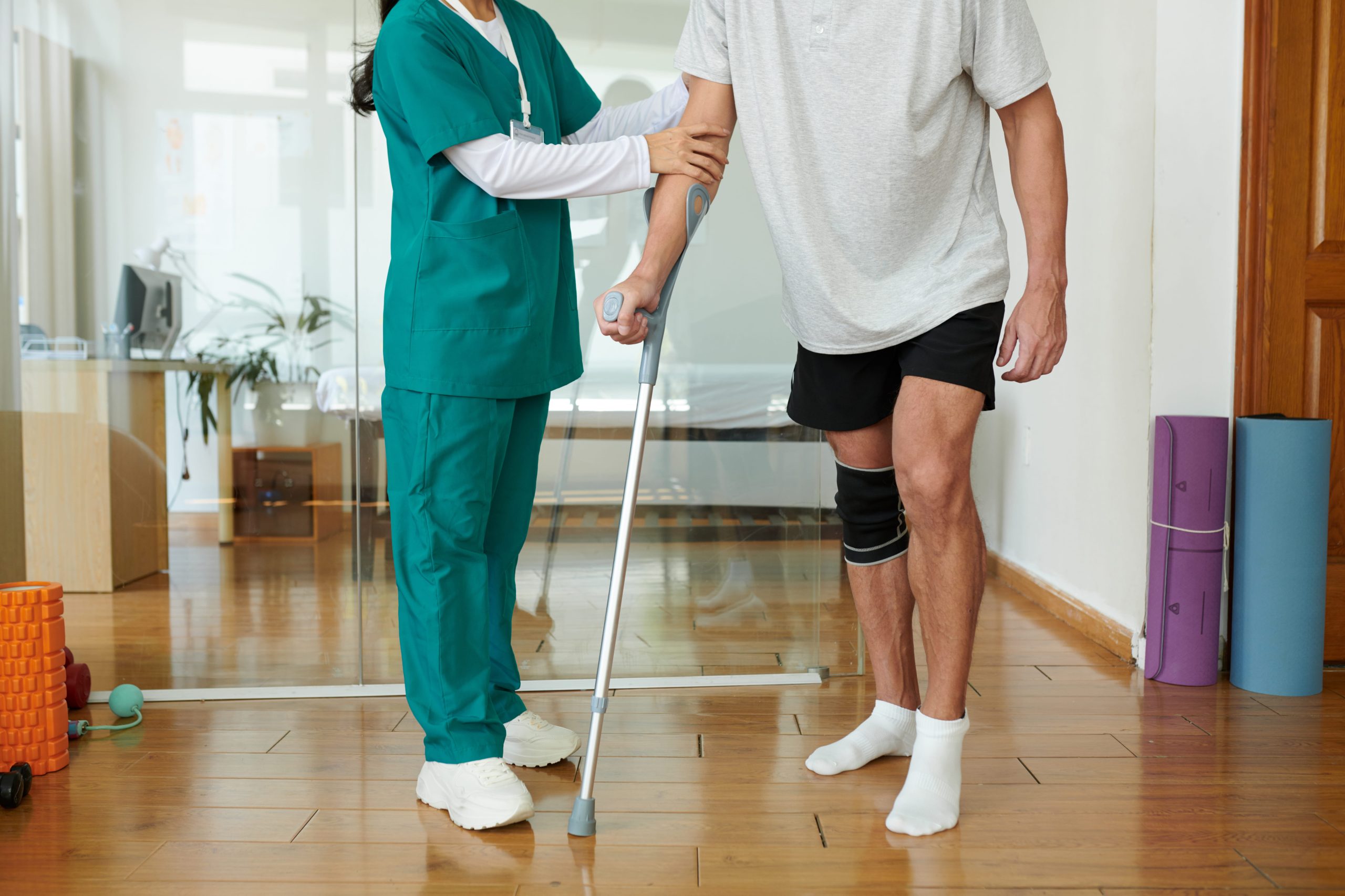
- Recovery: Medial meniscus repair typically requires protected weight-bearing for several weeks and restricted deep knee bending during healing, with return to full activity commonly occurring between four and six months following surgery, though individual timelines vary.
Treatment Approaches for Lateral Tears
Lateral meniscus treatment prioritises tissue preservation wherever possible, given the high proportion of load transmitted through this structure and the risk of accelerated compartment degeneration following removal.
- Repair as primary goal: Clinicians generally aim to pursue repair for lateral tears when anatomically feasible, as lateral meniscectomy, even partial removal, may accelerate lateral compartment degeneration more rapidly than equivalent medial procedures based on available long-term radiological data.
- Popliteal hiatus tears: Tears in the popliteal hiatus region may heal less predictably due to the absence of capsular attachment, and while specialised repair techniques address these posterior lateral tears, outcomes may vary when compared to repairs in other locations.
- Discoid meniscus: Lateral tears associated with discoid meniscus, an anatomical variant where the meniscus covers more of the tibial plateau than normal, may require saucerisation, which involves reshaping the meniscus to more typical dimensions while preserving the stable peripheral rim.
Recovery Expectations
Meniscectomy recovery generally involves a shorter timeline compared to repair recovery. Many patients return to desk work within one to two weeks and may resume light physical activity by four to six weeks following partial meniscectomy, though individual timelines vary.
Meniscal repair requires longer rehabilitation to protect the healing tissue. Weight-bearing restrictions typically last four to six weeks, with progressive loading thereafter. Deep squatting and pivoting activities are generally restricted for three to four months. Return to competitive sports commonly occurs between four and six months for many patients.
Factors that may influence recovery include:
- Tear location relative to the blood supply
- Patient age and tissue quality
- Concurrent injuries requiring treatment
- Pre-injury activity level and muscle conditioning
- Compliance with rehabilitation protocols
Physical therapy is an important component of treatment regardless of whether management is surgical or conservative. Quadriceps weakness can develop rapidly after a knee injury and may contribute to ongoing symptoms if not addressed.
Quick Tip: Maintaining quadriceps strength before any planned surgery may support post-operative recovery. Simple exercises such as straight leg raises and wall sits can typically be performed daily while awaiting treatment.
Long-Term Considerations
Meniscal tissue does not regenerate once removed. Each meniscectomy may increase contact pressure on the underlying cartilage, contributing to accelerated wear over time. This effect helps explain why preserving meniscal tissue through repair, when feasible, may support better long-term joint health than removal.
Activity modification may become necessary following significant meniscal loss. High-impact activities and deep squatting positions place greater stress on meniscal-deficient compartments. Many patients maintain active lifestyles by transitioning to lower-impact activities.
Weight management directly affects knee joint loading, and maintaining a healthy body weight may help reduce stress on both intact and repaired meniscal tissue.
When to Seek Professional Help
- Knee pain persists beyond two weeks after injury
- Swelling that recurs with activity
- Catching, locking, or giving way sensations
- Inability to fully straighten your knee
- Pain along the joint line with squatting
- Difficulty walking without a limp
- Popping or clicking accompanied by pain
Commonly Asked Questions
Can a meniscus tear heal without surgery?
Small tears in the outer vascular zone may respond to conservative management, including activity modification and physical therapy. Larger tears, tears in the avascular inner zone, and tears causing mechanical symptoms may typically require surgical intervention for resolution. Individual suitability depends on tear characteristics and a clinical assessment.
How do I know if my tear is medial or lateral?
Pain location provides the primary clue. Medial tears tend to cause inner knee pain, while lateral tears may produce outer knee pain. However, referred pain patterns and concurrent injuries can complicate self-assessment. Physical examination and MRI provide detailed clinical localisation.
Does a meniscus tear always require surgery?
Many meniscus tears, particularly degenerative tears and small stable tears, may respond well to non-surgical treatment. Surgery may become necessary when mechanical symptoms persist, when the knee locks, or when conservative measures fail to provide adequate relief.
What activities should I avoid with a meniscus tear?
Deep squatting, pivoting movements, and high-impact activities may aggravate meniscal symptoms. Specific restrictions depend on tear type and location. Your treating physician can provide individualised guidance based on your particular injury pattern.
Is medial or lateral meniscus repair more successful?
Both locations may achieve comparable repair outcomes when performed in appropriate candidates. Research suggests the lateral meniscus’s greater mobility can complicate posterior horn repairs, while the medial meniscus’s firm attachment provides more stable tissue for suturing. Patient age, tear pattern, and concurrent procedures are primary factors that influence outcomes alongside tear location alone.
Next Steps
Tear location, pattern, and patient factors help determine whether conservative management, repair, or partial meniscectomy may be appropriate. Early assessment through clinical examination and MRI may help prevent delays that could limit repair eligibility or allow secondary cartilage damage. A locked knee or persistent mechanical symptoms warrant prompt evaluation rather than a wait-and-see approach.
If you are experiencing inner or outer knee joint line pain, a catching or locking sensation, or difficulty fully straightening your knee, consult our accredited orthopaedic surgeon for a clinical assessment and to discuss your treatment options.