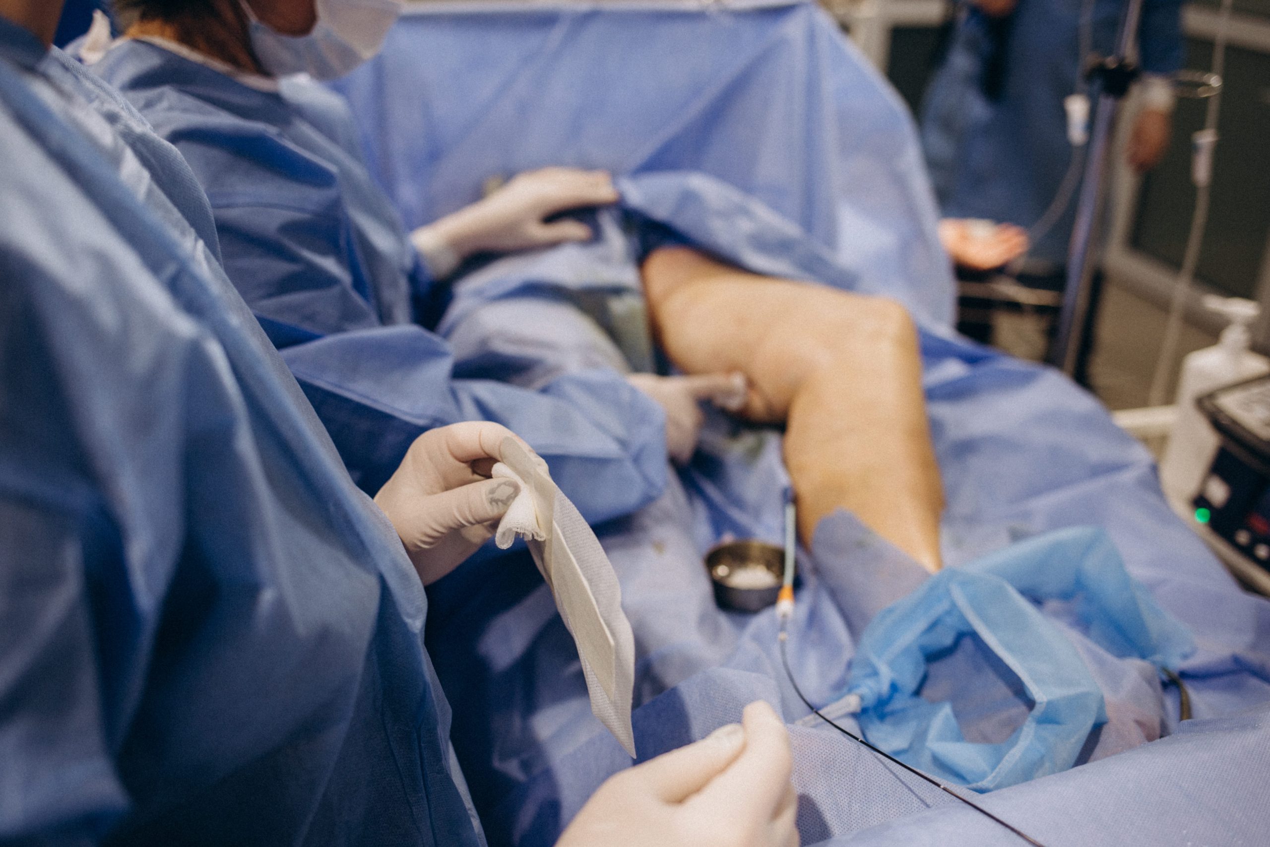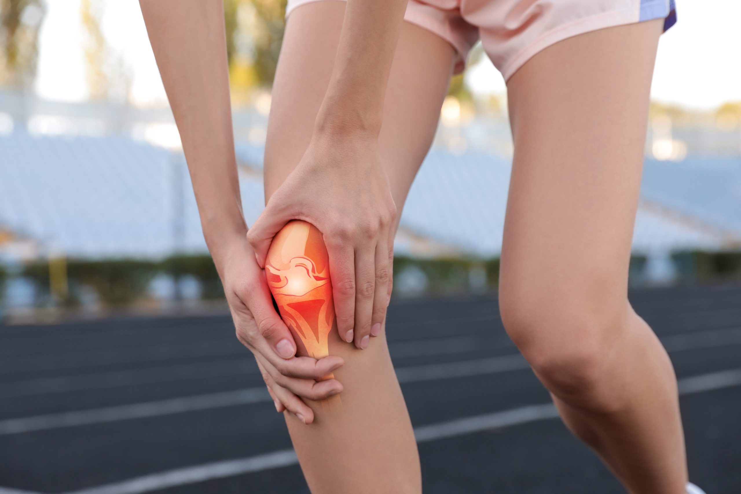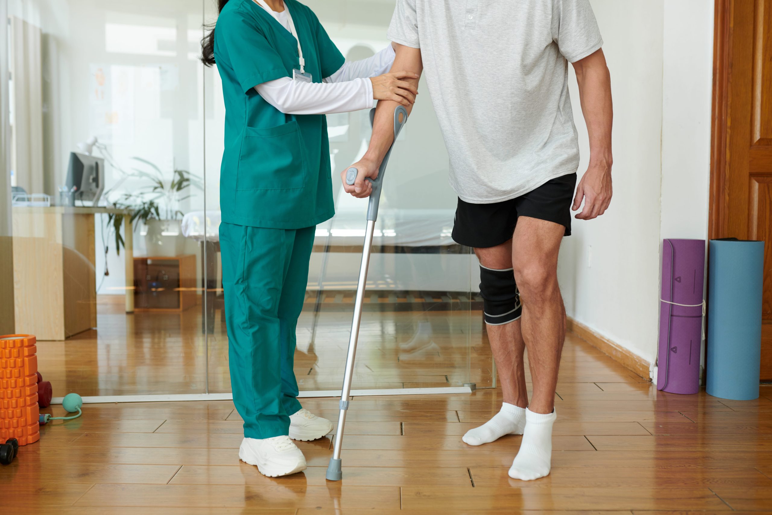
Graft choice in ACL reconstruction directly determines where your scars will be and how noticeable they become. The procedure typically requires multiple incisions: one or two small portal sites for arthroscopic instruments and a separate incision for harvesting the replacement graft.
Patellar tendon grafts require a vertical incision over the kneecap, while hamstring grafts produce a smaller incision along the inner shin, each with different aesthetic outcomes. Most ACL surgery scars transition from raised and reddened to flatter and lighter over 12 to 18 months, with continued fading possible for up to two years after surgery.
Incision Locations by Graft Type
Patellar Tendon (Bone-Tendon-Bone) Grafts
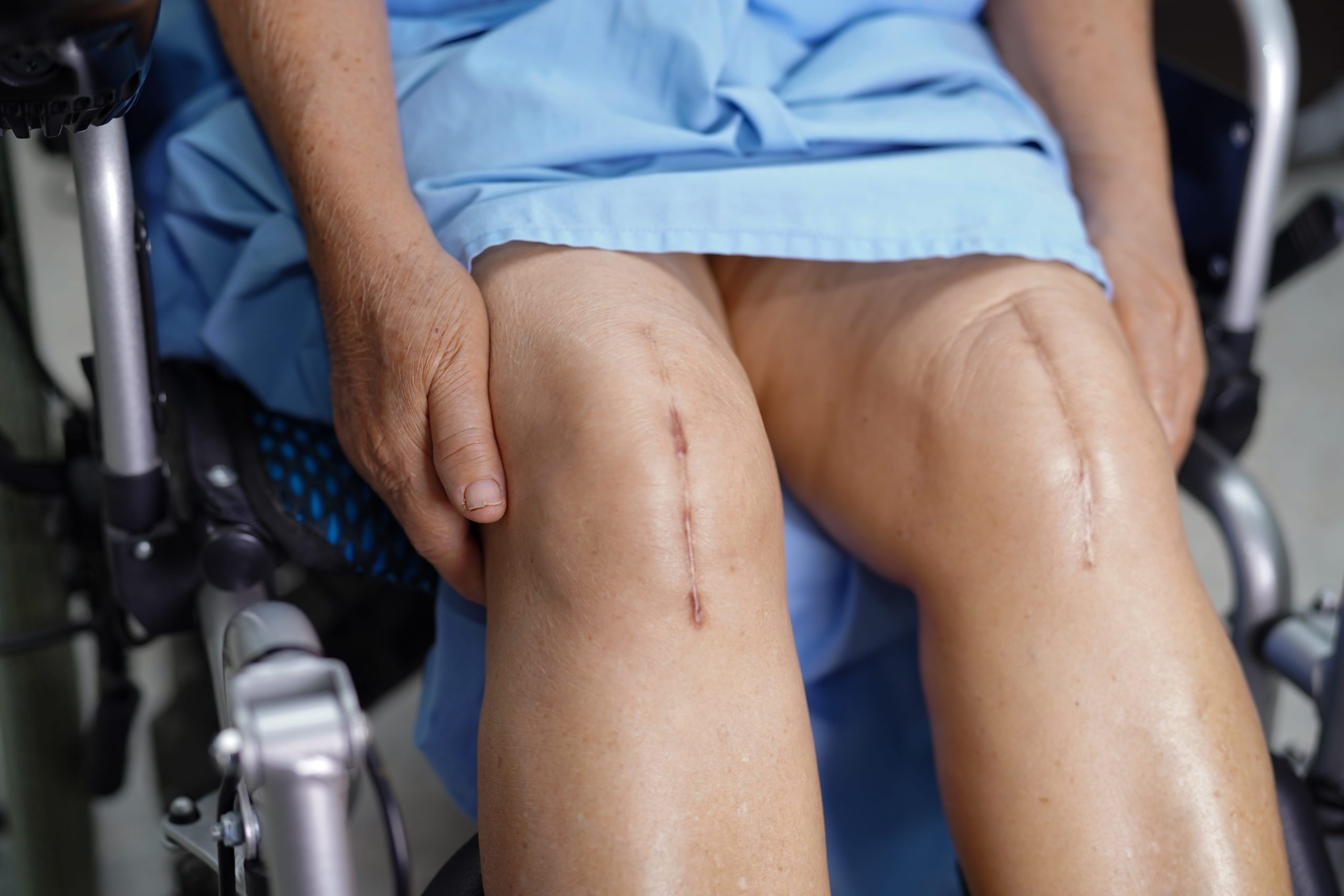
This technique requires a vertical incision running directly over the patellar tendon, typically measuring 6 to 8 centimetres. The incision extends from the lower portion of the kneecap to just below the tibial tubercle. Surgeons harvest the central third of the patellar tendon along with small bone plugs from both the kneecap and shinbone.
The location over the knee’s most prominent surface means this scar remains visible during activities like kneeling. Some patients report persistent tenderness at the harvest site, particularly when pressure is applied directly to the scar tissue.
Hamstring Tendon Grafts
Hamstring harvesting uses an oblique or vertical incision along the anteromedial tibia, positioned several centimetres below the joint line on the inner aspect of the shin. This incision typically measures 3 to 4 centimetres and sits in a less conspicuous location than patellar tendon scars.
The gracilis and semitendinosus tendons are extracted through this single incision. Because the harvest site sits along the inner leg rather than over the kneecap, many patients find this scar less noticeable during daily activities.
Quadriceps Tendon Grafts
A transverse or vertical incision above the kneecap provides access to the quadriceps tendon. This approach has gained popularity as an alternative graft source. The incision typically measures 4 to 5 centimetres and sits in the suprapatellar region, where clothing often covers the resulting scar.
Arthroscopic Portal Sites
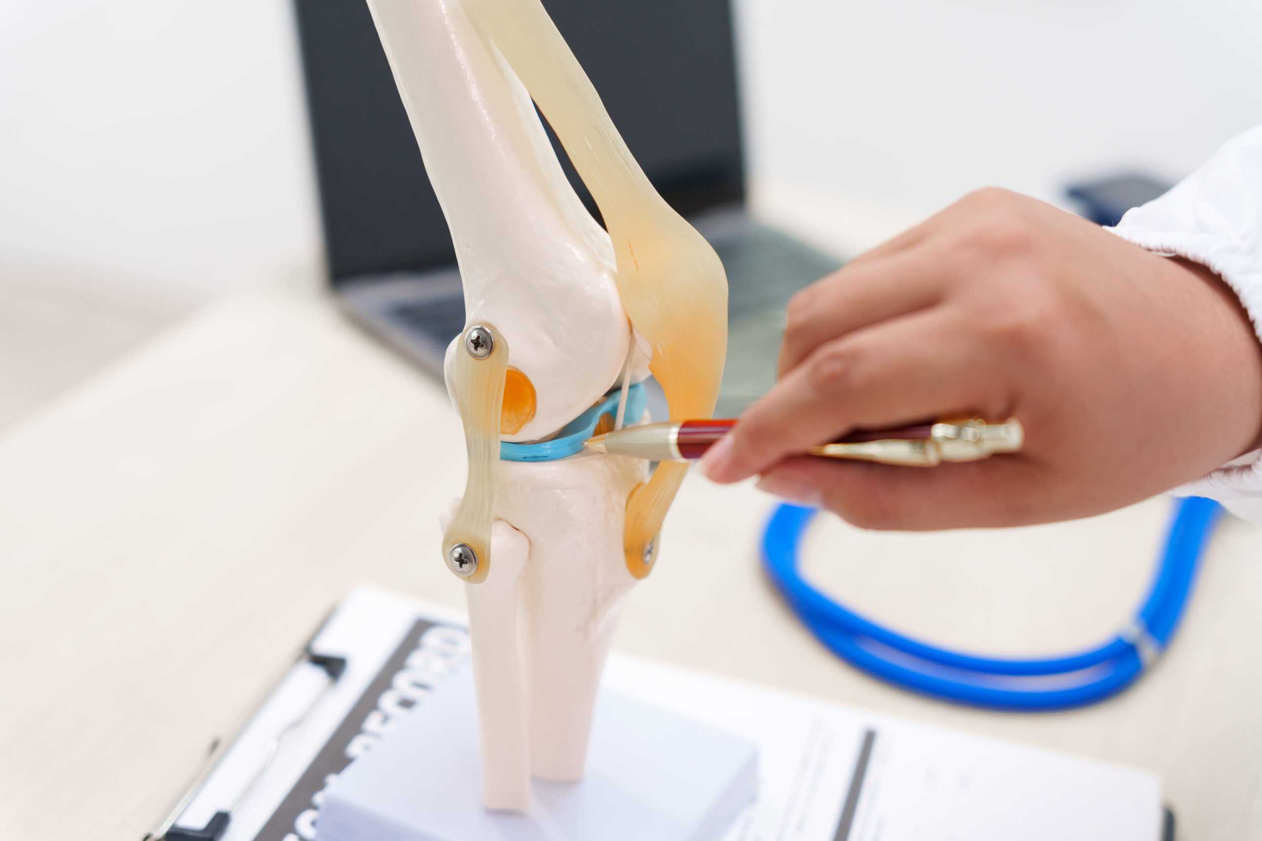
Regardless of graft choice, all ACL reconstructions require small arthroscopic portals. Two to three puncture-style incisions, each measuring 5 to 10 millimetres, allow camera and instrument insertion into the knee joint. These portal scars sit on either side of the patellar tendon and typically heal as small, circular marks that fade considerably over time.
The Biological Phases of Scar Healing
Inflammatory Phase (Days 1-7)
Wound healing begins immediately after incision closure. Blood clotting initiates the repair cascade, and inflammatory cells migrate to the wound site. Scars appear red and swollen and may feel warm to the touch. Bruising commonly extends beyond the incision lines during this phase.
Sutures or surgical staples maintain wound integrity while underlying tissue begins knitting together. Keeping incisions clean and dry during this phase reduces the risk of worsening eventual scarring.
Proliferative Phase (Weeks 2-6)
Collagen production accelerates as fibroblasts populate the wound bed. New blood vessels form within the healing tissue, contributing to the scar’s red or pink appearance. The wound gains tensile strength but remains vulnerable to stretching forces.
Scars often appear raised and firm during this phase. The body produces collagen abundantly, sometimes creating temporary thickness that later remodels. Gentle range-of-motion exercises prescribed during rehabilitation do not negatively affect scar formation when performed correctly.
Remodelling Phase (Months 2-24)
Collagen reorganises along lines of tension, and excess scar tissue gradually breaks down. The scar transitions from red to pink to white as blood vessel density decreases. Maximum scar maturation typically occurs between 12 and 18 months post-surgery, though subtle changes continue beyond this point.
Factors Affecting Scar Appearance
Skin Type and Genetics
Individuals with darker skin tones face a higher risk of keloid or hypertrophic scar formation. Family history of abnormal scarring predicts similar responses to surgical incisions. Those with known scarring tendencies should discuss preventive strategies with their surgeon before reconstruction.
Fair-skinned individuals may develop scars that initially appear more red or purple but often fade to near skin colour with time. Freckled or sun-damaged skin around the knee can create uneven scar pigmentation during healing.
Surgical Technique
Precise wound closure with appropriate tension minimises scar spreading. Layered closure techniques, where deeper tissues are sutured separately from skin, produce better cosmetic outcomes than single-layer closure. Attention to cosmetic detail influences final results.
Wound complications during initial healing, including infection, dehiscence, or haematoma, significantly worsen scarring. Meticulous post-operative wound care reduces these risks.
Age and Health Status
Younger patients generally heal with less visible scarring due to more robust collagen production and skin elasticity. However, younger individuals may also produce more aggressive scar tissue initially.
Smoking impairs wound healing through reduced oxygen delivery to tissues. Diabetic patients face prolonged healing times and increased infection risk. Nutritional deficiencies, particularly in protein, vitamin C, and zinc, compromise the body’s repair mechanisms.
Did You Know?
Scars never regain the full tensile strength of uninjured skin. Mature scar tissue reaches approximately 75 to 80% of the original skin strength, which explains why revision surgeries through previous scars require careful technique.
Evidence-Based Scar Management Techniques
Silicone-Based Products
Silicone sheets and gels represent the most studied scar treatment modality, with strong clinical evidence supporting their use for preventing and treating hypertrophic scars. Applied to healed wounds after suture removal and complete epithelialisation, silicone creates a hydrated environment that appears to regulate collagen production.
Sheets work best for flat scars on areas where adhesion is practical. Gels suit irregular surfaces or locations where sheets won’t stay in place. Consistent use for 12 to 24 hours daily over several months produces optimal results. Products should be applied to clean, dry skin without other creams or lotions underneath.
Massage Therapy
Scar massage mobilises tissue, breaks adhesions, and promotes collagen remodelling. Begin gentle massage once the wound has fully closed, typically two to three weeks after surgery. Apply firm pressure perpendicular to the scar line, working along its entire length.
Sessions of five to ten minutes, performed two to three times daily, provide therapeutic benefit. Use an unscented moisturiser to reduce friction. Massage also desensitises scars that remain tender to touch.
Sun Protection
Ultraviolet exposure causes permanent hyperpigmentation in healing scars. New scar tissue lacks melanin regulation, making it vulnerable to darkening that persists indefinitely. Cover scars with clothing or apply broad-spectrum sunscreen with SPF 30 or higher for the first year after surgery.
This precaution applies even on cloudy days and during brief outdoor exposure. Once a scar darkens from sun exposure, the discolouration rarely fades completely.
Compression
Pressure garments reduce hypertrophic scarring by limiting blood flow and collagen overproduction. While more commonly used for burn scars, compression can benefit surgical scars prone to thickening. Elastic bandages or fitted sleeves provide consistent pressure when worn for extended periods.
Managing Problematic Scars
Hypertrophic Scars
These raised, firm scars remain within the boundaries of the original incision. They typically develop within the first few months and may improve spontaneously over one to two years. Persistent hypertrophic scars respond to silicone therapy, compression, and intralesional corticosteroid injections administered by a medical professional.
Keloid Scars
Keloids extend beyond original wound margins and rarely regress spontaneously. They occur more frequently in individuals with previous keloid formation. Treatment options include corticosteroid injections, silicone therapy, pressure therapy, and occasionally surgical revision combined with adjuvant treatments to prevent recurrence.
Scar Contracture
Tight scar tissue can restrict knee flexion or extension if it forms across the joint line. Stretching exercises, prescribed as part of rehabilitation, help prevent contracture. Established contractures may require physiotherapy intervention or, rarely, surgical release.
Patients experiencing restricted range of motion should flag this promptly, as knee arthrofibrosis identified early responds better to conservative management than established contracture.
Important Note:
Redness, increasing pain, warmth, or discharge from a healing incision may indicate infection rather than normal scar maturation. These symptoms warrant prompt medical evaluation.
Timeline of Scar Appearance Changes
Weeks 1-2: Incisions appear red with visible sutures or staples. Swelling and bruising surround the wounds. The area feels tender and firm.
Weeks 3-6: After suture removal, scars may appear raised and remain red or dark pink. Itching commonly occurs as healing progresses. Early scar massage can begin.
Months 2-4: Scars reach peak firmness and may appear more prominent than they will long-term. Colour remains pink or red. Silicone therapy shows initial benefits.
Months 6-12: Gradual flattening and colour fading occur. Scars transition towards pink or light purple. Tenderness decreases significantly.
Months 12-24: Final maturation produces pale, flat scars in most patients. Some textural difference from the surrounding skin typically persists. Scars continue to subtly fade beyond this point.
Putting This Into Practice
- Begin silicone therapy promptly: Start within two weeks of suture removal and continue for three to six months.
- Protect from sun exposure: Cover scars or apply sunscreen daily for the first 12 months, regardless of season.
- Massage daily: Spend five to ten minutes on scar massage once wounds have fully healed, using firm perpendicular pressure.
- Stay hydrated and nourished: Adequate protein, vitamin C, and zinc support collagen production and wound healing.
- Attend follow-up appointments: Your surgeon monitors healing and can intervene early if problematic scarring develops.
When to Seek Professional Help
- Increasing redness, warmth, or swelling around incisions after the first week
- Discharge of pus or foul-smelling fluid from any wound
- Wound edges separating or sutures pulling through skin
- Scars are becoming increasingly raised or extending beyond the original incision lines
- Persistent pain at scar sites beyond three months
- Scar tissue restricting knee movement despite rehabilitation exercises
- Darkening or discolouration that concerns you
Commonly Asked Questions
Will my ACL surgery scars completely disappear?
Scars fade significantly but never disappear entirely. Most mature into pale, flat lines that blend reasonably well with surrounding skin. The arthroscopic portal scars often become barely noticeable, while graft harvest scars remain more visible due to their larger size.
When can I start treating my scars?
Begin scar management once wounds have fully closed and any sutures or staples have been removed, typically two to three weeks post-surgery. Starting silicone products and massage at this point allows intervention during the proliferative phase of healing.
Does the type of graft affect how visible my scars will be?
Yes. Hamstring grafts generally produce less conspicuous scarring because the harvest incision sits along the inner shin rather than over the kneecap. Patellar tendon grafts create a more visible vertical scar directly over the front of the knee. Discuss cosmetic considerations with your surgeon when reviewing graft options.
Can scar tissue inside the knee cause problems?
Internal scar tissue called arthrofibrosis can develop within the joint, limiting the range of motion. This differs from external skin scarring and requires specific rehabilitation protocols to prevent it itt. Diligent physiotherapy and early mobilisation reduce the risk of arthrofibrosis.
Should I consider scar revision surgery?
Most ACL surgery scars improve sufficiently without surgical intervention. Revision surgery is typically reserved for scars causing functional problems or those with severe cosmetic concerns that haven’t responded to conservative treatments after full maturation at 12 to 18 months.
Next Steps
Silicone therapy initiated within two weeks of suture removal, combined with daily sun protection for the first 12 months, yields the most documented improvement in the overall appearance of ACL surgery scars. The selection of a surgical graft fundamentally shapes final incision placement and long-term visibility, with hamstring harvests producing a less prominent harvest scar than patellar tendon approaches. Scars that become abnormally thickened, extend past the initial wound boundaries, or restrict active joint motion warrant professional evaluation before full natural maturation is reached at 18 months.
If you are managing an external surgical scar that is thickening, changing colour, or creating mechanical resistance during standard knee movement following an ACL reconstruction, a formal physical examination and tissue review by an accredited orthopaedic specialist can help clarify which scar management options align safely with your recovery.