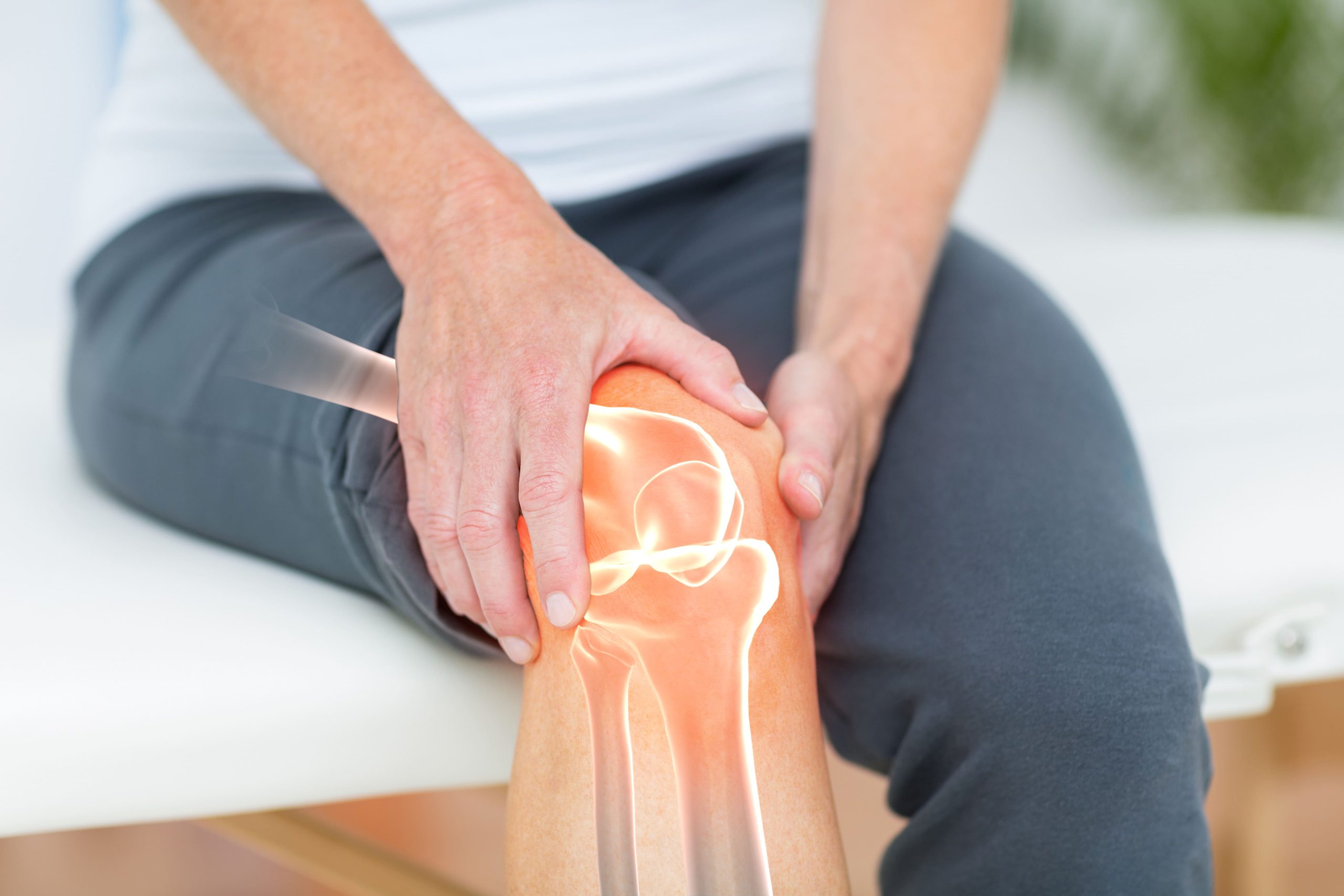
Articular cartilage lacks blood vessels. This severely limits its natural healing capacity. When damaged by injury or wear, significant cartilage defects, particularly those that do not reach the underlying bone, have limited self-repair capacity and typically progress over time rather than heal spontaneously.
Cartilage restoration surgery encompasses several techniques designed to regenerate or replace damaged cartilage before joint replacement becomes necessary. These procedures work by stimulating the body’s repair mechanisms, transplanting healthy cartilage tissue, or implanting laboratory-cultured cells to fill defects and restore smooth joint surfaces. The goal remains consistent across techniques: preserve native joint anatomy, restore pain-free movement, and delay or eliminate the need for artificial joint replacement.
How Cartilage Damage Occurs
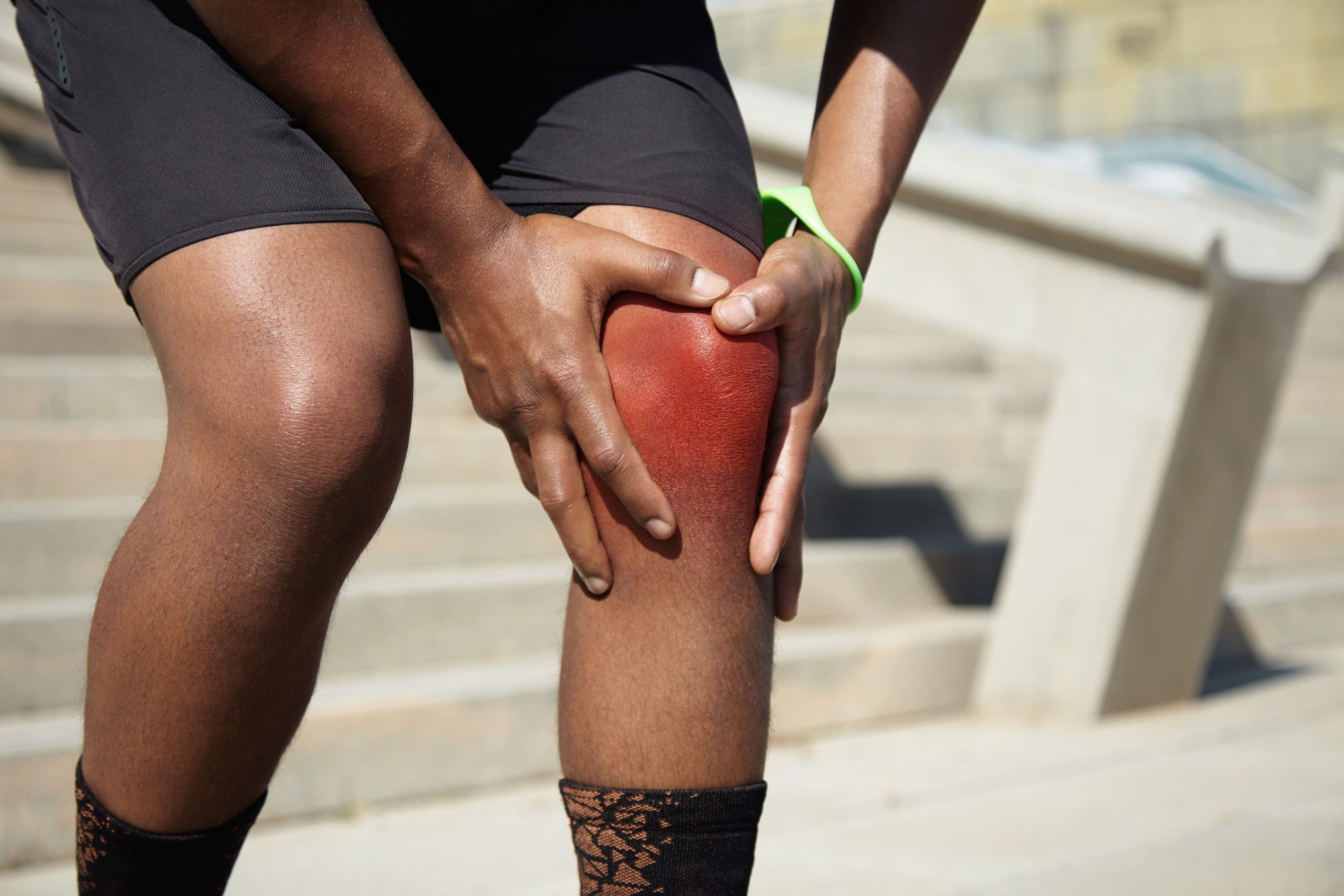
Cartilage injuries typically fall into two categories: traumatic and degenerative. Traumatic defects result from acute injuries, a pivoting movement during sports, a direct blow to the knee, or a fall that impacts the joint. These injuries often create focal lesions with well-defined borders in otherwise healthy cartilage. This makes them particularly suitable for restoration procedures.
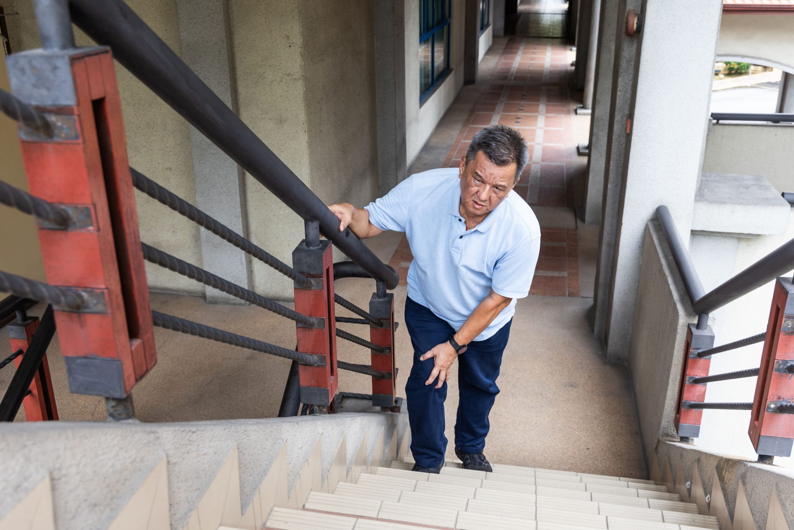
Degenerative damage develops gradually through repetitive stress, altered joint mechanics, or age-related changes. Previous ligament injuries that alter joint stability, meniscus tears that change load distribution, and limb malalignment all accelerate cartilage breakdown. Unlike traumatic defects, degenerative changes often affect broader areas and may involve multiple joint surfaces simultaneously.
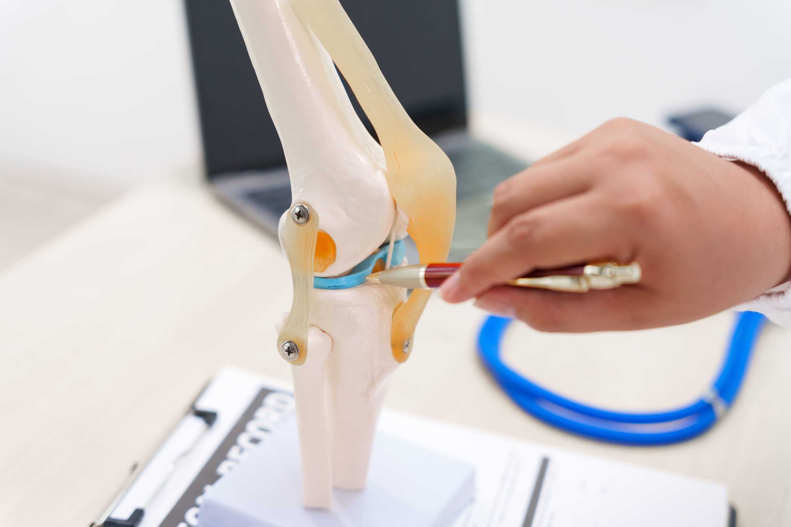
The International Cartilage Regeneration & Joint Preservation Society classifies cartilage damage by depth and extent:
- Grade I: Softening without surface disruption
- Grade II: Partial-thickness fissures
- Grade III: Defects extend to, but not through, the subchondral bone
- Grade IV: Lesions penetrate to bone, sometimes exposing or damaging the underlying surface
Your healthcare provider will select a treatment tailored to your specific classification, defect size and location, and individual risk factors.
Microfracture and Bone Marrow Stimulation
Microfracture remains a commonly performed cartilage restoration procedure due to its relatively straightforward technique and minimal equipment requirements. The surgeon creates multiple small holes through the damaged cartilage into the underlying bone. This allows bone marrow elements—including mesenchymal stem cells (cells that can develop into various tissue types) and growth factors (proteins that promote healing)—to fill the defect.
Procedure and Healing Process
Surgeons perform microfracture arthroscopically (using a small camera and instruments inserted through tiny incisions). The procedure involves first cleaning away the damaged cartilage to create stable, well-defined defect borders. Using a specialised awl (a pointed surgical tool), the surgeon penetrates the bone layer beneath the cartilage at 3-4mm intervals. This maintains spaces that allow structural stability whilst permitting marrow elements to escape. Blood and marrow form a clot within the defect. This clot gradually matures into fibrocartilage (repair tissue) over several months.
Clinical Considerations
Fibrocartilage differs structurally from native hyaline cartilage (the smooth, resilient cartilage normally found in joints)—it contains more type I collagen (a structural protein found in skin and tendons) rather than type II collagen (the main protein in joint cartilage). This makes it less resilient under repetitive loading. Clinical outcomes show significant early improvement in pain and function, though durability varies. Smaller defects (under 2-3 cm²), younger patients, and lesions in areas of lower mechanical demand tend towards more favourable results.
Rehabilitation requires protected weight-bearing for 6-8 weeks to allow the marrow clot to mature without disruption. Continuous passive motion (gentle machine-assisted joint movement) during this period appears to improve cartilage tissue quality. Most patients return to low-impact activities by 4-6 months, with gradual progression to higher-demand activities based on clinical progress.
Osteochondral Autograft Transfer
Osteochondral autograft transfer (OAT), also called mosaicplasty, involves harvesting cylindrical plugs of healthy cartilage with underlying bone from non-weight-bearing areas of the joint. Surgeons then transplant them into the defect. This technique transfers mature, hyaline cartilage rather than relying on repair tissue formation.
Surgical Technique
The surgeon harvests one or more cylindrical grafts from low-contact-pressure areas of the knee — typically the margins of the trochlea or the intercondylar notch — regions that bear minimal load during normal activities. These plugs, typically 6–8mm in diameter, are press-fit into corresponding holes created in the defect area.
Outcomes and Limitations
OAT provides immediate structural cartilage with proven durability in medium-term follow-up studies. The technique works well for defects generally between 1–2.5 cm², and may be considered for lesions up to 4 cm² in selected cases, particularly in the femoral condyles, where circular defects match the cylindrical graft geometry. Limitations include donor-site morbidity—harvesting creates new defects that fill with fibrocartilage—and difficulty in precisely matching curved joint surfaces. The technique is less suitable for defects in the patella (kneecap) or tibial plateau (top of the shin bone) due to geometric constraints.
Osteochondral Allograft Transplantation
When defect size exceeds what autograft can address, or when bone loss accompanies cartilage damage, osteochondral allograft transplantation offers an alternative. This technique uses donor tissue from deceased donors. This provides mature cartilage-bone units without creating additional harvest site defects.
Fresh allografts maintain cartilage cell viability; cryopreservation (freezing) significantly reduces chondrocyte (cartilage cell) survival. This makes tissue timing a logistical consideration. Healthcare providers typically aim to implant grafts as soon as disease testing is complete, generally from 15 days post-harvest, with 28 days representing the maximum recommended storage period to preserve adequate cellular function.
The procedure addresses defects ranging from several square centimetres to the entire joint surface. Surgeons can contour grafts to match the recipient geometry precisely. This makes the technique valuable for large traumatic defects, osteochondritis dissecans lesions (a condition in which bone and cartilage separate from the joint surface), and focal areas of cartilage loss. Integration of the bone portion occurs through gradual creeping substitution (a process where the transplanted bone is slowly replaced by the patient’s own bone), whilst the cartilage surface maintains its structure throughout healing.
Autologous Chondrocyte Implantation
Autologous chondrocyte implantation (ACI) is an established cell-based cartilage restoration technique performed in two surgical stages:
- Initial arthroscopic harvest of healthy cartilage cells, which are then sent for laboratory expansion over several weeks
- Subsequent implantation of the expanded cells into the prepared defect
First-Generation Technique
Original ACI secured a periosteal patch (tissue harvested from the tibia or femur) over the defect. This created a sealed chamber into which expanded chondrocytes were injected. Whilst effective, this approach required open surgery and carried risks of periosteal hypertrophy (excessive tissue growth) requiring secondary procedures.
Matrix-Assisted Techniques
Current approaches embed cultured chondrocytes within collagen or synthetic scaffolds (supporting structures that help guide tissue growth) before implantation. These MACI procedures are typically delivered via mini-arthrotomy, with arthroscopic delivery emerging as an option for accessible defects, reducing surgical morbidity and providing structural support during tissue maturation.
Patient Selection
ACI and MACI are suitable for larger defects (typically over 2-3 cm²) where simpler techniques show diminishing effectiveness. The procedures require intact surrounding cartilage borders, stable joint mechanics, and patient commitment to extended rehabilitation. Concurrent procedures addressing ligament instability or limb malalignment can improve outcomes by optimising the mechanical environment for healing tissue.
Emerging Cell-Based Approaches
Research continues into alternative cell sources and delivery mechanisms with the hope of developing improved techniques. Mesenchymal stem cells (cells that can develop into various tissue types, including cartilage) harvested from bone marrow or adipose tissue (fat) offer theoretical advantages—they avoid the need for cartilage biopsy and may have broader regenerative potential. Clinical evidence supporting these approaches is still in its early stages compared to established techniques.
Particulated juvenile allograft cartilage uses minced donor tissue from donors typically aged 13 years or younger. This provides chondrocytes with higher proliferative capacity than adult cells.
However, clinical evidence for this approach remains at an earlier stage than established techniques, and short-term follow-up data have reported complication rates, including graft hypertrophy, and a modest failure rate.
Factors Affecting Procedure Selection
Selecting the appropriate cartilage restoration approach requires evaluating multiple patient and defect characteristics. Your surgeon will consider defect size, location, depth, and chronicity, along with your age, activity demands, body mass index, and joint alignment, to establish goals tailored to your individual risk factors and health needs.
Defect Characteristics
Small, contained lesions with healthy surrounding cartilage often respond well to microfracture or OAT. Larger defects typically require cell-based approaches or allograft transplantation. Defects involving significant bone loss necessitate techniques that address both layers—allograft transplantation or staged bone grafting followed by surface restoration.
Location matters significantly. Weight-bearing femoral condyle lesions see good results across techniques. Patellar and trochlear defects present challenges due to high shear forces and complex geometry. Tibial plateau lesions require careful consideration of the softer underlying bone.
Patient Factors
Younger patients with acute traumatic defects generally achieve better outcomes than older patients with degenerative changes. Activity expectations influence technique selection—an athlete may accept longer rehabilitation for potentially more durable repair, whilst a less active patient might prefer simpler approaches with faster recovery.
Body mass places increased demands on healing cartilage. Patients with elevated body mass index may be advised by their surgeon to consider weight management before surgery, as higher body weight places increased demands on healing cartilage and has been associated with less favourable outcomes in some studies. However, the decision to proceed with or delay surgery should be made in consultation with a qualified specialist, taking into account the individual’s overall health.
Complementary Procedures
Cartilage restoration rarely occurs in isolation. Many patients with cartilage defects have concurrent pathology that contributes to their condition or results from the same injury.
Addressing Underlying Causes
Anterior cruciate ligament reconstruction (surgical repair to restore knee stability) may precede or accompany cartilage restoration when instability threatens the repair tissue. Meniscal repair or transplantation restores load distribution across the joint surface. High tibial osteotomy or distal femoral osteotomy (procedures that realign the leg bones) corrects limb malalignment. This shifts the load away from damaged compartments.
Addressing these factors prevents the mechanical environment that caused initial cartilage damage from destroying restoration efforts. Staged procedures, correcting alignment first, then performing cartilage restoration, may produce better outcomes than single-stage combined approaches in selected cases.
Rehabilitation Principles
Post-operative rehabilitation follows consistent principles across cartilage restoration techniques, though specific protocols vary by procedure, defect location, and surgeon preference.
Healthcare providers begin early, almost immediately, to promote cartilage nutrition and prevent adhesions (scar tissue that limits movement). Continuous passive motion machines or frequent range-of-motion exercises maintain joint mobility whilst protecting healing tissue from excessive force.
Weight-bearing restrictions allow repair tissue to mature without disruptive compressive loads. Most protocols progress from non-weight-bearing or toe-touch weight-bearing to partial, then full, weight-bearing over 6-12 weeks. Progression timing depends on the procedure performed, defect size and location, and the radiographic or clinical assessment of healing.
Strengthening focuses initially on non-weight-bearing exercises. This progresses to closed-chain activities (exercises in which the foot remains in contact with the ground, such as squats) as weight-bearing increases. Impact activities and sports-specific training occur in later rehabilitation phases, typically 6-12 months post-operatively, depending on the procedure.
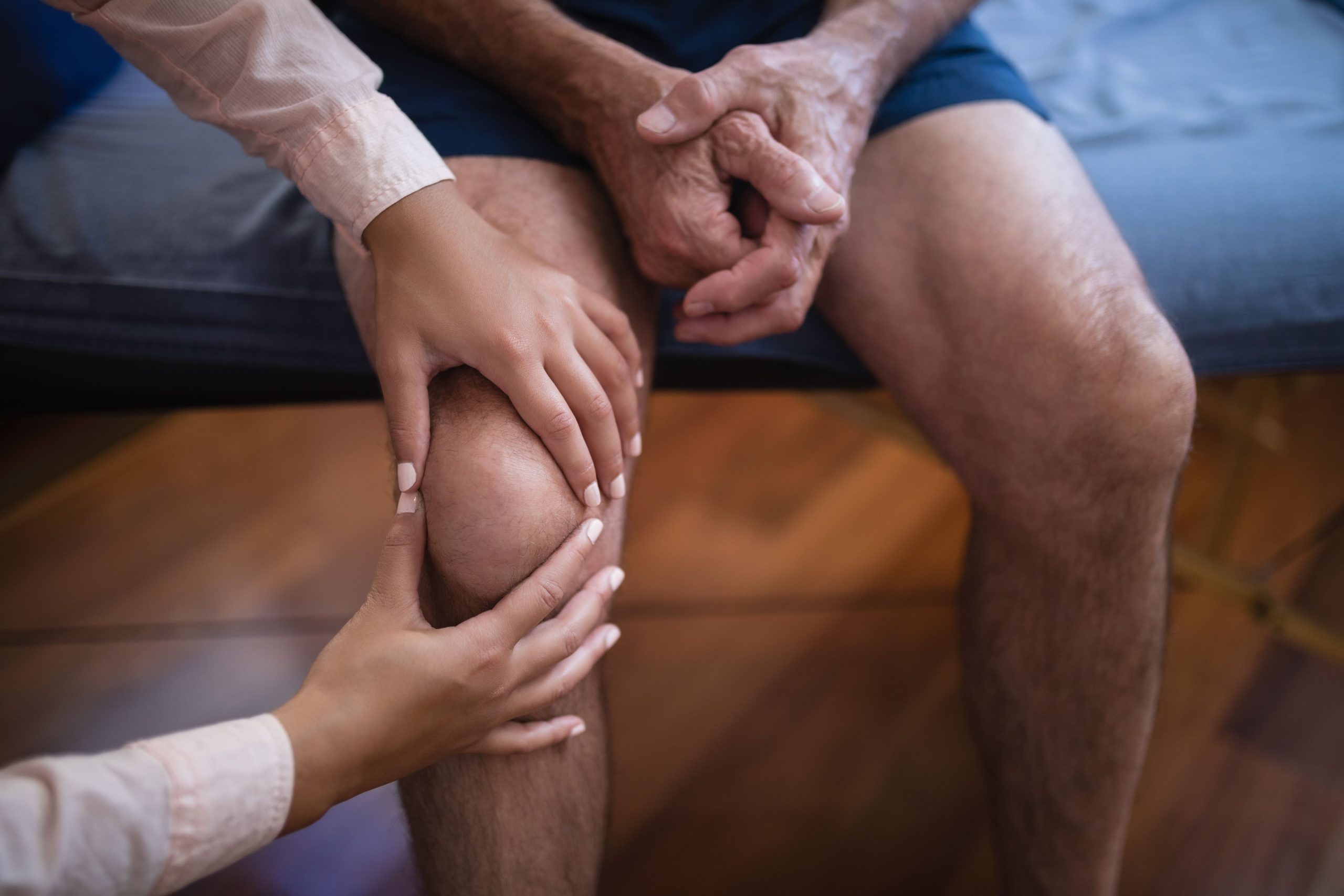
When to Seek Professional Help
- Persistent joint pain that interferes with daily activities or sleep
- Swelling that recurs with activity or doesn’t resolve with rest
- Mechanical symptoms such as catching, locking, or giving way
- Progressive difficulty with activities you previously performed without discomfort
- Joint pain following a specific injury that doesn’t improve over several weeks
- Stiffness following periods of activity or prolonged inactivity
Commonly Asked Questions
How long does recovery take after cartilage restoration surgery?
Microfracture typically allows a return to daily activities within 3 to 4 months for smaller lesions, though full recovery generally takes 6–12 months. Return to sport averages approximately 8 months in athletes. Cell-based procedures and allograft transplantation often require 9–12 months before returning to demanding activities. Complete tissue maturation continues for 1–2 years after surgery.
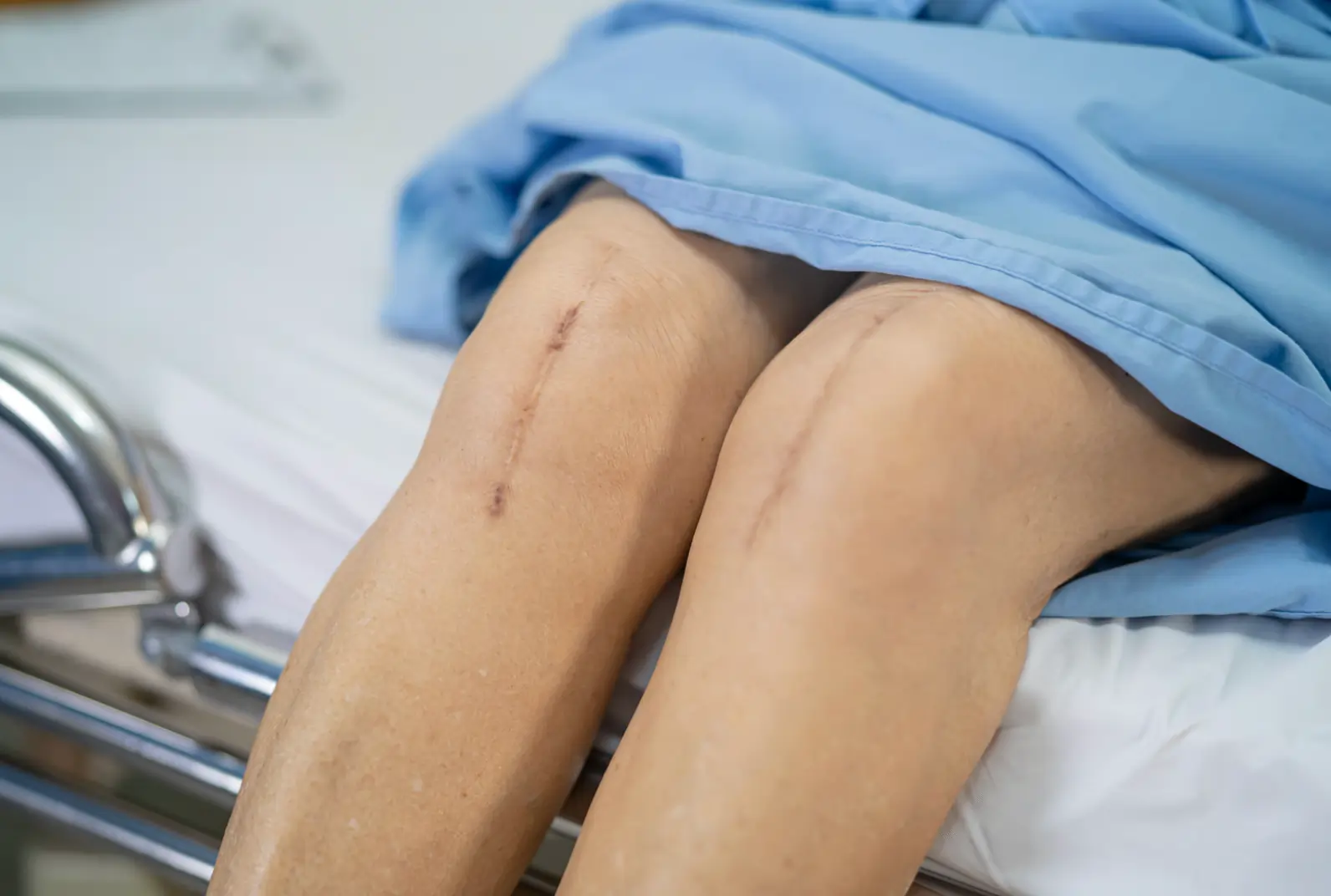
Can cartilage restoration prevent the need for joint replacement?
When performed in appropriate candidates, cartilage restoration procedures can preserve native joint function for many years. The goal is to delay or eliminate the need for joint replacement, particularly in younger patients for whom artificial joints have a limited lifespan.
What happens if cartilage restoration fails?
Healthcare providers can often revise failed cartilage restoration with alternative techniques. A failed microfracture may be followed by cell-based therapy or allograft transplantation. Surgeons may revise failed cell-based procedures with repeat cell implantation or an allograft. When restoration options are exhausted or inappropriate, partial or total joint replacement remains available.
Are the results of cartilage restoration permanent?
Restored cartilage tissue demonstrates durability in clinical studies, though long-term outcomes vary by technique and patient factors. Repair tissue quality, patient activity level, body weight, and joint mechanics all influence longevity. Some patients maintain good function indefinitely, whilst others experience gradual deterioration requiring additional intervention.
What activities can I return to after cartilage restoration?
Most patients return to recreational activities, including cycling, swimming, golf, and hiking. The return to high-impact sports depends on the procedure performed, the defect location, and the healing quality. Running and cutting sports are possible for some patients but carry a higher risk of reinjury or deterioration of repaired tissue.
Next Steps
Untreated cartilage defects progress over time, and early intervention offers the opportunity to preserve native joint function. Procedure selection depends on defect size and location, patient age, activity demands, and the presence of concurrent joint pathology such as ligament instability or malalignment. Addressing these factors concurrently is often necessary to protect any restoration performed.
If you are experiencing persistent joint pain, swelling, or mechanical symptoms such as catching, locking, or giving way, consult a qualified orthopaedic specialist for a formal evaluation of your cartilage health.